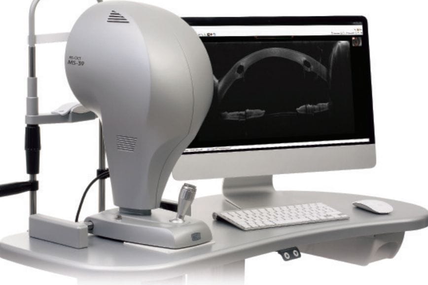
The revolutionary MS-39 is a highly flexible anterior segment topographer-tomographer that can replace numerous instruments in your surgical practice. It has been shown to enhance patient flow, and management of corneal, refractive, cataract and other surgical procedures.
The MS-39 combines placido disk corneal topography with high resolution optical coherence tomography (OCT) based anterior segment tomography.
The list of possibilities is quite staggering…
 This sophisticated anterior segment topographer-tomographer provides more detailed information than traditional Scheimpflug cameras, with faster capture times. But this is just the beginning – the versatile MS-39 is far more than a new generation topographer. Additional features facilitate high resolution OCT imaging, infra-red pupillography, tear film analysis, lens biometry, and intraocular lens (IOL) calculations. Coupled with the Phoenix software program, it is a powerful tool that can replace numerous other instruments, and improve patient flow by providing faster scanning and analysis.
ANTERIOR SEGMENT OCT
The standard anterior segment OCT topographer scans in up to 25 meridians over a 16mm area of the eye. This provides data for 12 different corneal maps, including anterior and posterior curvature and elevation, refractive power, corneal pachymetry and epithelial thickness. Analysis also shows keratometry readings for anterior, posterior and total corneal power, as well as metrics for horizontal visible iris diameter, thinnest and central pachymetry, anterior chamber and corneal volume. This comprehensive suite of data can be acquired in just one second per eye. A separate aberrometry module shows maps and tables of higher order aberrations.
HIGH DEFINITION ANTERIOR SEGMENT OCT IMAGING
In addition to routine scans, the MS-39 can capture high resolution images of the anterior ocular structures, including anterior and posterior corneal surfaces, the iris and the anterior chamber angle. The software then accurately measures corneal thickness, epithelial thickness, anterior chamber depth, and angle geometry, using a tool known as a ‘magic wand’. The magic wand automatically measures the relevant distance as you hover the cursor over the area. Separate tools allow manual measurements in any location. This is useful for quantifying unusual features, like visualising and measuring the depth and location of corneal scarring. It can also accurately measure the location and positioning of a phakic IOL after surgery, or document and quantify the fit of rigid corneal and scleral contact lenses.
 Screening Modules
Phoenix software includes tailored modules to screen and monitor keratoconus and glaucoma. The keratoconus module allows early and accurate detection of corneal ectasia, with significant statistical analysis and classification of severity. A keratoconus monitoring report allows comparison with previous scans for detailed monitoring.
The glaucoma module combines corneal pachymetry and anterior chamber data to calculate angle opening distance and the trabecular-iris space area. This provides additional information and tools for glaucoma diagnosis and management.
IOL Power
When combined with axial length measurements, this module uses ray tracing techniques to calculate both spherical and toric IOL powers. Axial length measuring capability is expected in future upgrades.
Lens Biometry
A detailed biometric scan provides analysis of the crystalline lens thickness and position, as well as information about lens tilt and anterior chamber depth.
Pupillography
Infra-red pupillography measures pupil size in scotopic, mesopic and photopic illumination. There is also a dynamic mode which captures video analysis of pupil movement under variable illumination levels.
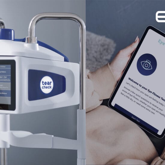
Tear Film Analysis
Placido disk technology allows for advanced analysis of tear break up time. A clever video capture system starts automatically when the patient blinks. The timing and location of tear break up over time is shown in map and graph formats.
Phoenix Software
The software package includes a typical patient database, with examinations saved in chronological order and labelled according to scan type for fast and easy comparison and review. Display and print out of corneal maps is completely customisable – users can specify colour schemes, step sizes, the number and type of maps, and other statistics to appear on print outs. There is also a huge database of examples, with reference scans from patients with dozens of different ocular conditions.
EXPERIENCE IN THE REAL WORLD
We have had the MS-39 in our busy refractive surgery practice for a number of months. The instrument and accompanying software are very user friendly, both for the operator and patient. One of the stand out features is the amount of data that can be obtained in a very short space of time – what the MS-39 obtains in its one second standard tomography scan (corneal tomography, epithelial thickness and anterior chamber measurements) would otherwise require numerous measurements on at least two separate instruments. This improves patient flow through the practice, as moving patients between instruments is fiddly and time consuming.
Comparison with other instruments
By bringing together the features of high definition OCT and placido disk, the MS-39 is able to perform everything that a Scheimpflug tomographer provides, but the faster capture time makes it easier to achieve good quality scans, which reduces the need for repeated scanning. Additionally, its OCT imaging capabilities are simpler and faster than obtaining the equivalent information on epithelial thickness or anterior chamber parameters using traditional OCT.
Surgery Planning and Follow Up
When used to its full potential, the MS-39 can revolutionise the way a practice manages the planning and follow up of corneal, refractive, cataract and other surgical procedures. This instrument can be used to obtain most, or all, of the initial measurements, perform IOL calculations, and then quantify and monitor changes following surgery.
 The list of possibilities is quite staggering: surgically induced astigmatism, epithelial thickness variation after corneal laser, position and stability of phakic IOLs, anterior chamber angle changes following glaucoma procedures, corneal monitoring with collagen crosslinking, the various corneal transplant modalities, and so on. The unique combination of high resolution images and precision tomography enable the surgeon to both visualise and quantify anterior segment abnormalities, changes, and surgical outcomes.
Dr Peter Stewart is the Director of Surgery at Lasersight. He graduated from the University of Queensland in 1973 and began general medical practice in far western Queensland. Following an inspirational meeting with Professor Fred Hollows, Dr Stewart began his training in ophthalmology in Sydney.