


An evolving understanding of polypoidal choroidal vasculopathy (PCV), as a subtype of wet age-related macular degeneration (nAMD), is changing the way clinicians approach treatment and management of this disease, which affects far more people than previously realised.
Dr Voraporn Chaikitmongkol from Chiang Mai University Hospital in Thailand, and Professor Gemmy Cheung from the Singapore National Eye Centre, presented the latest research on PCV at Bayer’s Global Retinal Network Program for Australia and the Asia Pacific in 2021.
PCV is a chronic eye disease in which fluid leaks and bleeding occurs from abnormal blood vessels of the choroid.
Dr Chaikitmongkol’s research has demonstrated that colour fundus photography (CFP), which is widely available and noninvasive, has a high sensitivity and specificity for identifying PCV when used in conjunction with optical coherence tomography (OCT)

Dr Voraporn Chaikitmongkol.
A subtype of nAMD, the prevalence of PCV is much higher in Asian than Caucasian populations. While the prevalence of PCV among clinic patients presenting with exudative AMD was believed to be 4-15% in Caucasian populations and 25-78% in Asian eyes, Dr Voraporn Chaikitmongkol, a retinal specialist from Thailand, says the prevalence is likely to be much higher.
In a convincing presentation, Dr Chaikitmongkol explained that indocyanine green angiography (ICGA) has been accepted as the gold standard for diagnosis of PCV, however ICGA is invasive and time consuming. Furthermore, in Thailand, 66% of retinal centres do not have ICGA and similarly, other countries in the region have limited access to ICGA. This means many cases of PCV are going undiagnosed.
She said alternative imaging modalities are now shining the light on the true prevalence of this potentially blinding condition.
Dr Chaikitmongkol’s research has demonstrated that colour fundus photography (CFP), which is widely available and non-invasive, has a high sensitivity and specificity for identifying PCV when used in conjunction with optical coherence tomography (OCT).
Clinical clues to diagnose PCV on CFP are:
- Subretinal orange-red nodules,
- Fibrovascular pigment epithelialdetachment (PED),
- Massive subretinal haemorrhage,
- Peripapillary location, and
- No large drusen in the fellow eye.
Important to note is that PCV eyes can present with or without drusen. So, if a patient presents with a sub macular haemorrhage in one eye, and drusen (hallmark of early staged AMD) in the fellow eye, they might have advanced nAMD or PCV.
If a patient presents with a sub macular haemorrhage in one eye, and no drusen in the fellow eye, it is more likely to be PCV, or diseases other than nAMD.
Even when used alone, Dr Chaikitmongkol asserted that OCT has high sensitivity and specificity for PCV detection when compared to ICGA.
PCV characteristics on OCT are:
- Notched PED,
- Peaked PED,
- Hyper reflective ring underneath PED,and
- The ‘double layer sign’ – a thinseparation between retinal pigmentepithelium and Bruch’s membrane – which is the appearance of branching vascular network.
In 2019, Dr Chaikitmongkol’s group published a paper in JAMA Ophthalmology: Sensitivity and specificity of potential diagnostic features detected using fundus photography, optical coherence tomography, and fluorescein angiography for polypoidal choroidal vasculopathy.1
In other words, because of limited access to ICGA, they had set out to determine whether fundus photography, optical coherence tomography, and fluorescein angiography would be as accurate in diagnosing PCV as ICGA.
This retrospective study reviewed imaging of 120 patients with newly diagnosed serous or serosanguinous maculopathy. Patients underwent four types of imaging, between 1 January 2013 and 31 December 2016, at Chiang Mai University Hospital in Thailand.
Dr Chaikitmongkol’s group found that FFA had a low level of accuracy when identifying PCV and concluded that for clinicians with OCT and CFP, there is no need to use FFA.
When using OCT and CFP, her group concluded that it is necessary to identify at least two of the following four features to diagnose PCV with 93% accuracy:
- Presence of notched or haemorrhagicPED or fibrovascular PED on CFP,
- Presence of sharply peaked non-serousPED with an angle of 70-90 degreeson OCT scan,
- Notched or multilobulated non-serousPED seen on OCT scan, and
- Presence of hyperreflective ringunderneath the PED – of any size.
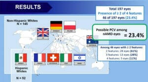
Dr Chaikitmongkol’s research, using non ICGA diagnostic criteria, found the prevalence of probable PCV diagnosis among nAMD eyes in Caucasians is more likely 23.4% – significantly higher than the number reported in the literature (4%-14%), and closer to the 22% reported by Dr Gerald Liew in his paper, Prevalence of polypoidal choroidal vasculopathy in Caucasian patients from optical coherence tomography signs.2
TREATMENT OUTCOMES
Dr Chaikitmongkol moved on to describe real world research which has confirmed clinical studies showing that PCV is highly responsive to treatment with aflibercept monotherapy.
The primary aim when treating PCV is to improve vision. Additionally, anatomical outcomes sought through treatment include:
- Complete polypoidal regression,
- Reduction in the number of polypoidallesions and polypoidal lesion area, and/or
- Polypoidal lesion inactivation.
The PLANET study found after one year of treatment with aflibercept monotherapy, the mean visual acuity improvement was >10 letters from baseline.
In a study titled Timing of complete polypoidal regression following intravitreous aflibercept treatments in polypoidal choroidal vasculopathy,3 Dr Chaikitmongkol’s group followed 40 eyes in Thailand with PCV that each received three, monthly injections of aflibercept followed by injections every eight weeks for one year. They found that at one year, 55% had complete polypoidal regression on ICGA; 40% had partial regression and just 5% had no regression. Of the eyes with complete regression at one year, 50% achieved complete regression at two months after aflibercept initiation and 77% had achieved complete regression within the first six months.
While the available data from this study did not enable Dr Chaikitmongkol’s group to determine whether it is possible to stop treatment when complete regression is achieved, her continued research aims to determine this.
Delving into the detail, she said the PLANET study has shown that monotherapy can achieve excellent visual and anatomical outcomes, comparable to initial PDT with anti-VEGF
PCV AND EXTENDED TREATMENT INTERVALS
Professor Gemmy Cheung continued the discussion on treatment of PCV, exploring evidence for achieving extended treatment intervals and whether this disease should be treated differently from nAMD.
She proposed that our understanding of PCV as a subtype of nAMD (as opposed to a specific entity) has changed the approach to its treatment – from occlusive/destructive to control of activity; from PDT/laser therapy to anti-VEGF monotherapy, and from a reactive approach to a proactive approach.
Prevalence of PCV in Caucasian populations is likely to be significantly higher than the numbers reported in the literature.
Delving into the detail, she said the PLANET study has shown that monotherapy can achieve excellent visual and anatomical outcomes, comparable to initial PDT with anti-VEGF.
The PLANET study also showed that following the first year of treatment, dosing could be reduced from 8.1 to 4.6 treatments of aflibercept monotherapy between week 52 and 96, while still retaining excellent outcomes.
The ALTAIR study further investigated the potential to extend treatment intervals in patients with nAMD, with either two or four week treatment interval adjustments, and tolerating some residual subretinal fluid in the absence of other disease activity criteria. On average, vision gains were achieved and maintained through week 96 for patients with and without PCV in this study. A large proportion of patients achieved 12- and 16-week intervals, and these outcomes have been supported by real world studies.
Professor Cheung led a study comparing aflibercept with a personalised treat and extend (T&E) regimen vs. fixed bi-monthly dosing for PCV, which was performed in the Singapore National Eye Centre.4
They found the vision gains at week 52 in the personalised treat-and-extend arm were non-inferior to those in the fixed arm. Additionally, they used used ICGA findings at week 12 to guide the personalised treatment.
Following three four-weekly loading doses, patients in the T&E arm with persistent polypoidal lesion on ICGA, regardless of subretinal or intraretinal fluid, received a further three four-weekly aflibercept injections before the T&E regimen was started. Patients in the T&E arm in whom the polypoidal lesion has closed on ICGA could start T&E as early as week 12.
Prof Cheung reported that 69% of patients in the personalised T&E arm achieved treatment intervals of more than or equal to 10 weeks at week 52.
At week 12, 40% of patients in the personalised T&E arm achieved complete closure of polypoidal lesions, increasing to 55% at week 24. This rate was maintained at 55.2% at week 52.
This compared favourably against the bimonthly arm, in which 46.2% of patients achieved closure at week 12. The closure rate remained largely unchanged and was 41.6% at 52 weeks.
Prof Cheung recommended clinicians start with monotherapy, with a loading phase of three four-weekly aflibercept injections, before evaluating for polypoidal lesions with ICGA at week 12. If lesions are present, administer a further course of three four-weekly injections before moving to a T&E regimen. If there are no polypoidal lesions at 12 weeks, proceed with a T&E regimen. She said this individualised protocol provides optimal treatment for all patients with PCV, ensuring residual polypoidal lesions are addressed while reducing retreatment burden during the T&E phase. Additionally, it allows patients with a low treatment need to extend treatment intervals at an early stage.
In closing, Prof Cheung reasserted that the efficacy of aflibercept for PCV has been established in clinical trials. However, many patients continue to need treatment beyond two years. A T&E approach should be considered to balance maintaining vision against retreatment burden and to achieve a higher polyp closure rate, additional loading or deferred PDT may be considered.
Prof Cheung said even in eyes that have achieved polypoidal lesion closure, new polypoidal lesions can present, and old lesions can reoccur, leading to bleeding or exudation. The optimal monitoring interval and imaging modality of these eyes remain unclear.
Long-term treatment data will help guide us on the need for continued treatment following achievement of polyp closure. Continued vigilance and proactive treatment is necessary to mitigate the risk of exudation or polypoidal lesions recurring.
Look forward to Bayer’s next GRNP this year.
Hero image: Professor Gemmy Cheung.
References
- Chaikitmongkol V, Kong J, Khunsongkiet P, Patikulsila D, Sachdeva M, Chavengsaksongkram P, Dejkriengkraikul C, Winaikosol P, Choovuthayakorn J, Watanachai N, Kunavisarut P, Ingviya T, Bressler NM. Sensitivity and Specificity of Potential Diagnostic Features Detected Using Fundus Photography, Optical Coherence Tomography, and Fluorescein Angiography for Polypoidal Choroidal Vasculopathy. JAMA Ophthalmol. 2019 Jun 1;137(6):661- 667. doi: 10.1001/jamaophthalmol.2019.0565. PMID: 30973593; PMCID: PMC6567979.
- Liew G, Hyun-Jin HD, Hooper C, Chia EM, Mitchell P, Ong S, Ho IV. Prevalence of polypoidal choroidal vasculopathy in Caucasian patients as estimated from optical coherence tomography signs. Eye (Lond). 2021 Mar;35(3):1011-1012. doi: 10.1038/s41433-020-0834-z. Epub 2020 Mar 9. PMID: 32152515; PMCID: PMC8027611.
- Chaikitmongkol V, Upaphong P, Patikulsila D, Jirarattanasopa P, Choovuthayakorn J, Watanachai N, Kunavisarut P, Ratanasukon M, Bhurayanontachai P, Ingviya T, Bressler SB, Bressler NM. Timing of Complete Polypoidal Regression after Intravitreous Aflibercept Treatments in Polypoidal Choroidal Vasculopathy. Ophthalmol Retina. 2021 Mar 27:S2468-6530(21)00101-9. doi: 10.1016/j. oret.2021.03.012. Epub ahead of print. PMID: 33781929.
- Teo KYC, Jordan-Yu JM, Tan ACS, et al. Efficacy of a novel personalised aflibercept monotherapy regimen based on polypoidal lesion closure in participants with polypoidal choroidal vasculopathy. British Journal of Ophthalmology Published Online First: 11 February 2021. doi: 10.1136/ bjophthalmol-2020-318354


