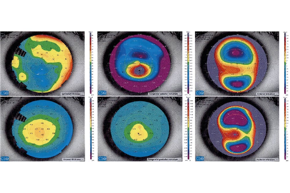
Figure 1. Customised MS39, six map display anterior segment OCT corneal mapping.
Technology is par excellence, as are patients’ refractive outcomes and expectations. As Drs Erica Darian-Smith and Mitchell Lee write, refractive surgeons have an extensive armamentarium of diagnostic equipment to help them make informed decisions regarding treatment suitability, surgical plans, and ultimately to predict outcomes, including epithelial coherence tomography (OCT) mapping.
One of the critical centre points in planning refractive surgery is epithelial optical coherence tomography (OCT) mapping – a noninvasive imaging technique that allows for detailed visualisation and measurements of the corneal epithelium. OCT is based on low coherence interferometry, which uses near-infrared light to produce high-resolution images of the layered corneal tissue morphology.1
CORNEAL EPITHELIUM: WHY IS IT SO IMPORTANT?
The corneal epithelium is approximately 55 μm in thickness and is generally thinner superiorly (by approximately 6 μm) due to the upper lid eyelid tension. When the underlying stroma is irregular, the overlying epithelium remodels to compensate for these changes. This plasticity and compensatory property of the epithelium can be seen in various conditions such as: epithelial basement membrane dystrophy (EBMD), keratoconus, post corneal surgeries, and corneal scars.
DIFFERENTIATING MASQUERADING CONDITIONS
When using topography in isolation, it can sometimes be difficult to differentiate contact lens warpage (CLW) from keratoconus (KCN) because both exhibit inferior steepening. Epithelial OCT imaging can be beneficial to differentiate the two conditions when a patient has early keratoconus changes. Inferior thickening of the epithelium is seen in CLW whereas KCN demonstrates the characteristic thinning over the cone and compensatory surrounding donut shaped epithelial thickening.2
ROLE IN DIAGNOSING ECTASIA AND MEDICOLEGAL IMPLICATIONS
The corneal epithelium has very characteristic changes in cases of keratoconus or corneal ectasia. There is a classical thinning of the epithelium over the apex of the cone and compensatory thickening in a ‘donut’ appearance around it. Reinstein et al. first reported this classical configuration seen in keratoconus cases.3 Of note, very early subclinical cases of keratoconus can be detected by using epithelial mapping. It is of utmost importance to identify these cases so that surgeons can ensure refractive surgery is not performed; this group is at a high risk of developing post-laser ectasia. This provides an additional layer of medicolegal protection, which is important in the litigious landscape that we reside in. Patients who have early keratoconus detected can also benefit from ongoing surveillance and potentially, treatment with corneal collagen cross-linking early if there are features of progression.
AVAILABLE DEVICES AND REPRODUCIBILITY OF RESULTS
There are many different devices on the market that measure epithelial thickness. These include but are not limited to:
- MS39 anterior segment OCT and tomographer (CSO, Italy),
- Avanti OCT System (Optovue, United States),
- Spectralis OCT (Heidelberg, Germany),
- Cirrus HD-OCT (Carl Zeiss Meditec, Germany), and
- Insight 100 VHF (very high frequency) digital ultrasound scanner (ArcScan, United States).
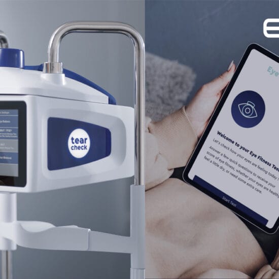
At our practice, Eagle Eye Surgeons Laser Vision Correction, we use the MS39 device. This uses spectral-domain (SD) OCT combined with Placido disc corneal topography and has a resolution of 3.6 μm in tissue. The stromal and epithelial thickness can be calculated over an 8 mm diameter. For reproducibility as well as ease of diagnosis of ectasia, we use the Reinstein suggested and customised six map keratoconus summary page split into the following: 1. Epithelial thickness, 2. Tangential anterior, 3. Anterior elevation, 4. Stromal thickness, 5. Tangential posterior, and 6. Posterior elevation (Figure 1).
Several studies have shown high repeatability of epithelial thickness measurements using this device.4,5 When comparing epithelial thickness map results between Anterion, MS-39 and Avanti, the thickest values were with the MS-39, followed by the Avanti and Anterion. Interestingly the differences in measurement of epithelial thickness between the devices means that the devices are not interchangeable when monitoring epithelial maps in patients with keratoconus.6,7 As clinicians we must choose our diagnostic machine of choice and stick with it to appropriately monitor progression for patients and also to compare inter-patient results within the practice.
PATTERNS OF EPITHELIAL THICKNESS CHANGES
It is useful to understand the patterns of epithelial thickness change in commonly encountered refractive surgery scenarios.
Pre-operative
Pattern recognition of epithelial thickness changes, for a variety of commonly encountered conditions and pathologies, is crucial during the patient selection process for laser surgery to ensure efficacy and safety of the procedure.2
- Post orthokeratology use: Central thinning and mid-peripheral thickening.
- Epithelial-basement membrane disease: Thickening centrally and inferiorly.
- Dry eye: Superior epithelium is more prone to be thin and damaged with a greater range of map standard deviation.
- Ectatic or keratoconic cases: Classical donut pattern of epithelial thinning over the ectatic cone surrounded by an annulus of thicker epithelium.2
Post-operative
Remodeling of the corneal epithelium post laser surgery is dependent on a number of factors including ablation depth, optical zone size, type of refractive error, and type of procedure (lenticular extraction versus flap-based procedure).
- Post-myopic laser-assisted in situ keratomileusis (LASIK): Thickens across the central 6 mm with maximal thickening centrally. A greater ablation depth corresponds with more thickening centrally, and increasing the diameter of the optical zone decreases the amount of central epithelial thickening. Myopic regression correlates with an increase in central epithelial thickness.8,9
- Post-hyperopic LASIK: Thins centrally (2 μm of thinning for every diopter of hyperopic treatment) and thickens paracentrally. Thinning over the apex can occur with high hyperopic treatments and can be an indicator of the development of apical syndrome.10
- Post-myopic small incision lenticule extraction (SMILE): Epithelial thickness profile changes may impact on the refractive outcome in the long-term postoperative period. For instance central epithelial thickening was associated with regression in patients with high myopia (SE > -8D).11
KERATOCONUS SCREENING: MS39 VS PENTCAM
When comparing the Pentacam HR and the MS-39 for screening and monitoring keratoconus, the former measures steeper K2 values and also thinner pachymetry results compared with the MS-39. This means that the instruments are not interchangeable when monitoring progress and that diagnosing keratoconus progression should be based on the stage and the test re-test repeatability rather than a fixed K2 or pachymetry value.12
FINAL THOUGHTS
It is clear that epithelial OCT mapping has many useful indications, including identifying subclinical corneal ectasia, customising refractive surgery type based on epithelial regularity and thickness, and identifying underlying corneal conditions and masqueraders such as EBMD, which can affect refractive outcomes. By becoming proficient with interpreting epithelial OCT thickness, we can provide our patients with more comprehensive assessments to improve refractive outcomes, and have greater confidence when selecting and appropriately managing our patients.
Dr Erica Darian-Smith MBBS MMed GradDipRefCatSurg FRANZCO is an ophthalmologist who specialises in refractive, cataract, and corneal surgery. She is a Director at Eagle Eye Surgeons in Mosman, in Sydney.

Dr Mitchell Lee BSc MBBS (Hons) MMed(Critical Care) FRANZCO is an ophthalmologist who specialises in vitreoretinal surgery, as well as complex anterior segment, cataract, and lens surgery. He is a Director at Eagle Eye Surgeons in Mosman, Sydney.

References
- Huang D, Swanson EA, Lin CP, et al. Optical coherence tomography. Science 1991;254(5035):1178-81. doi: 10.1126/science.1957169.
- Abtahi M, Beheshtnejad AH, Abtahi SH, et al. Corneal epithelial thickness mapping: A major review. J Ophthalmol. 2024:6674747. doi: 10.1155/2024/6674747.
- Reinstein D, Srivannaboon S, Gobbe M, et al. Epithelial thickness profile changes induced by myopic LASIK as measured by Artemis very high-frequency digital ultrasound. J Refract Surg 2009;25(5):444-450. doi: 10.3928/1081597X-20090422-07.
- Schiano-Lomoriello D, Bono V, Abicca I, Savini G. Repeatability of anterior segment measurements by optical coherence tomography combined with Placido disk corneal topography in eyes with keratoconus. Sci Rep 2020;10:1124. doi: 10.1038/s41598-020-57926-7.
- Vega-Estrada A, Mimouni M, Espla E, et al. . Corneal epithelial thickness intrasubject repeatability and its relation with visual limitation in keratoconus. Am J Ophthalmol. 2019;200:255-262. doi: 10.1016/j. ajo.2019.01.015.
- Khamar P, Rao K, Wadia, K, Dalal R, et al. Advanced epithelial mapping for refractive surgery Indian J Ophthal. 2020;68(12):2819-2830. doi: 10.4103/ijo. IJO_2399_20.
- Feng Y, Reinstein DZ, Stokanovic A, et al. Epithelial thickness mapping in keratoconic corneas: Repeatability and agreement between CSO MS-39, Heidelberg Anterion, and Optovue Avanti OCT devices. J Refract Surg 2023;39(7):474-480. doi: 10.3928/1081597X- 20230606-01.
- Lohmann C, Guell JL. Regression after LASIK for the treatment of myopia: The role of the corneal epithelium. Semin Ophthalmol 1998;13(2):79-82. doi: 10.3109/08820539809059822.
- Reinstein DZ, Archer TJ, Gobbe, M. Rate of change of curvature of the corneal stromal surface drives epithelial compensatory changes and remodeling. J Refract Surg 2014;30(12):799-802. doi: 10.3928/1081597X- 20141113-02.
- Reinstein DZ, Archer TJ, Gobbe M. Epithelial thickness after hyperopic LASIK: three-dimensional display with Artemis very high-frequency digital ultrasound. J Refract Surg 2010;26(8):555-564. doi: 10.3928/1081597X- 20091105-02.
- Ganesh S, Brar S, Relekar KJ. Epithelial thickness profile changes following small incision refractive lenticule extraction (SMILE) for myopia and myopic astigmatism. J Refract Surg 2016;32(7):473-482. doi: 10.3928/1081597X-20160512-01.
- Seiler T, Mueller, M, Mendes Baiao, T. Repeatability and comparison of corneal tomography in mild to severe keratoconus between the anterior segment OCT MS-39 and Pentacam HR. J Refract Surg 2022;38:250-255.