
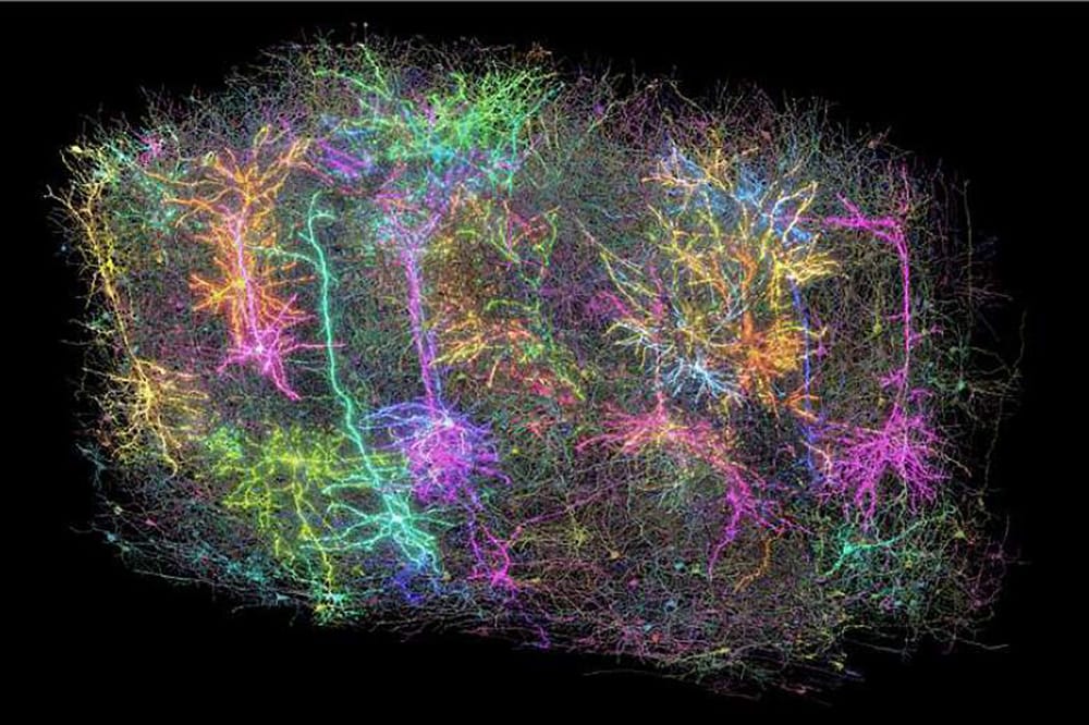
This image shows a subset of more than 1,000 of the 120,000 brain cells reconstructed in the MICRONS project. Each reconstructed neuron is a different random colour. It is meant as a symbolic representation of the dataset. There are far more recorded neurons than those that are glowing, and far more reconstructed neurons than were put into the rendering. Allen Institute.
Scientists are one step closer to understanding how the human brain processes visual information to reconstruct images in everyday life, having completed a massive scientific effort funded by the National Institutes of Health (NIH).
The acquired knowledge, which was previously beyond our reach, could help us understand how the brain functions normally and offer a guide to what goes wrong as the result of various disorders or injuries.
The acquired knowledge… could help us understand how the brain functions normally and offer a guide to what goes wrong as the result of various disorders or injuries
Complex Connections Revealed
The effort involved hundreds of researchers helping to map the connections between hundreds of thousands of neurons in the mouse brain and then overlaying their firing patterns in response to visual stimuli.
Information processing in the human brain occurs via electrical firing of 86 billion neurons that make trillions of connections with each other. The secrets of how our brain enable us to think, feel, and act lie hidden in the complexity of its wiring diagram and the barrage of electrical signals that move across it in millisecond time frames. While the current findings focus on a tiny fraction of the brain, they reveal the complex connections between the cells and show how those connections are wired to produce functional responses.
To carry out the study, researchers presented video clips to mice genetically engineered for their neurons to emit light when they fire. The neuron firing patterns in areas on the brain surface that are associated with vision were optically recorded across a cubic millimeter – about the size of a grain of sand. Within this deceptively small amount of tissue lies remarkable complexity: four kilometres of axons, the processes that nerve cells use to communicate with each other, intertwined to make more than 524 million connections called synapses across more than 200,000 cells.
Mapping Connections
To map these connections, teams worked 12-hour shifts for 12 straight days to carefully cut and image ultra-thin slices of the brain tissue using electron microscopes (EM). Reconstruction was the most challenging next step, as it required accurate stitching together almost 28,000 EM images to align the connections that cross the volume of brain tissue. This was followed by months of tracing the connections using deep learning algorithms followed by manual, and automated proofreading. Deep learning predictive models that explain visual information processing in the cortex were constructed and validated. In total, the sheer amount of data collected to create this tiny map comes out to 1.6 petabytes, roughly the equivalent of 22 years of continuous HD video.
These results come at a time when maps of neurons and their connections are increasingly revealing the mysteries of the brain. In 2023, research funded by the National Institutes of Health Brain Research Through Advancing Innovative Neurotechnologies Initiative, or The NIH BRAIN Initiative, produced the first complete cell atlas of the mouse brain, including the types and locations surveyed from more than 32 million cells. Last year, the NIH BRAIN Initiative ‘Flywire’ project led to the complete mapping of the common fruit fly brain, demonstrating the unique value of mapping the whole brain in its entirety.
Funding for this project was provided through the Machine Intelligence from Cortical Networks (MICrONS) Program of the Intelligence Advanced Research Projects Activity and the NIH BRAIN Initiative. The findings, published in a package of 10 papers published in the Nature family of journals, represent more than seven years of work performed by more than 150 scientists around the world.
The mouse connectome data detailed here can be visualised online using the MICrONS Explorer resource.


