In this comprehensive CPD-accredited review, Dr Nathan Kerr examines the latest diagnostic technologies, medical and surgical therapies, and emerging treatments that will define glaucoma care over the coming decade.
The landscape of glaucoma management has undergone a paradigm shift. We’ve moved beyond purely intraocular pressure (IOP)-centric treatment toward an integrated model combining earlier intervention, sustained drug delivery, and personalised risk stratification. For Australian optometrists and ophthalmologists, understanding these advances is critical – not only because an estimated 50% of glaucoma in Australia remains undiagnosed,1,2 but because collaborative care models are reshaping how we detect, treat, and monitor this sight-threatening disease.
Diagnostic Advances Reshaping Early Detection
Imaging technology has evolved beyond simple structural measurements to encompass vascular assessment, artificial intelligence (AI)-powered analysis, and home-based monitoring. These developments are fundamentally changing diagnosis and how we identify disease progression.
Swept-Source OCT and OCT-Angiography
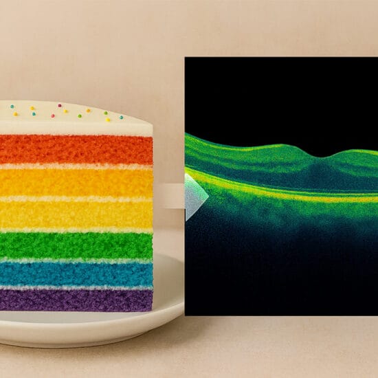
Swept-source optical coherence tomography (OCT) and OCT-angiography (OCT-A) now provide superior visualisation of deep optic nerve structures, including the lamina cribrosa, with particular advantages in myopic eyes where spectral-domain OCT can struggle. OCT-A is emerging as a powerful complementary tool, identifying microvascular damage in the optic nerve head and detecting capillary dropout in superficial and deep retinal layers.
Critically, vessel density measurements have lower measurement floors than retinal nerve fibre layer (RNFL) thickness – making them especially valuable for monitoring advanced disease where traditional OCT parameters plateau.3 Research indicates that retinal and choroidal vascular impairments occur during early glaucoma stages, potentially allowing detection before structural loss becomes apparent.
Artificial Intelligence
AI is transforming diagnostic accuracy, though important caveats remain. Deep learning algorithms analysing OCT achieve area under the receiver operating characteristic curve (AUC) values of 0.90–0.92 for distinguishing glaucomatous from healthy eyes, while fundus image-based systems report diagnostic accuracy in the mid-90% range in selected datasets.4 AUC quantifies a test’s ability to distinguish between disease and non-disease states, with values above 0.90 generally considered excellent.
Deep learning models analysing macular OCT can now identify patients experiencing rapid visual field loss (worse than −1 dB/year) – those most at risk of significant vision impairment – with AUC of 0.92.5 Explainable AI tools provide transparent, interpretable insights with AUC of 0.96–1.00 for severity staging.
However, no Therapeutic Goods Administration (TGA)-approved autonomous AI systems exist specifically for glaucoma screening, and current AI augments, rather than replaces, clinical judgement. Eye care professionals should view these tools as sophisticated assistants, not autonomous decision makers.
Home Intraocular Pressure Monitoring
Home intraocular pressure (IOP) monitoring has matured considerably. The iCare Home2 demonstrates excellent agreement with Goldmann tonometry (correlation coefficient 0.99, mean difference approximately 2 mmHg).6,7 Studies reveal that a majority of patients have IOP peaks outside typical clinic hours,8 making home monitoring valuable for patients progressing despite apparently controlled in-office pressures.
When diurnal or 24-hour monitoring is employed, treatment changes are made in around one-third to three-quarters of patients.9 This highlights how much traditional clinic measurements can miss, and underscores the value of home monitoring in appropriate candidates.
Virtual reality and home perimetry represents perhaps the most practice-changing development for Australian optometry.
Virtual Reality and Home Perimetry
Virtual reality and home perimetry represents perhaps the most practice-changing development for Australian optometry. The Melbourne Rapid Fields (MRF) system – developed at the University of Melbourne and TGA-registered – enables tablet-based and home visual field testing with validation showing high correlation to Humphrey Field Analyser (HFA) results (intraclass correlation coefficient 0.93 for mean deviation).10,11
Similarly, Eyeonic – a TGA-approved, cloud-based online perimetry platform also developed in Melbourne – enables visual field testing on any computer with internet access. Validation studies demonstrate diagnostic accuracy comparable to standard automated perimetry, with strong patient preference over conventional testing.
Home monitoring studies demonstrate high short-term uptake (around 85–90%) for weekly testing,12 and modelling indicates MRF testing every two months offers equivalent diagnostic power for detecting progression as six-monthly HFA Swedish Interactive Threshold Algorithm (SITA)-Standard in simulation models.13 These Australian innovations are enabling rural and outreach services, and fundamentally altering monitoring paradigms.
A United States Food and Drug Administration (FDA)-cleared wearable virtual reality platform also shows strong agreement with HFA in mild-to-severe glaucoma, with mean deviation correlation around 0.9 and high concordance for disease staging.14
Home monitoring studies demonstrate high short-term uptake (around 85–90%) for weekly testing
Polygenic Risk Scoring: SightScore
Genetic risk stratification represents one of the most significant advances in personalised glaucoma care. SightScore, developed by Australian researchers at Flinders University and QIMR Berghofer Medical Research Institute, and commercialised by Adelaide-based Seonix Bio, is the first clinically available polygenic risk score (PRS) for primary open-angle glaucoma (POAG).
The foundational research, published in Nature Genetics in 2020, analysed over 400,000 individuals and demonstrated remarkable predictive power.15 Individuals in the top PRS decile reach an absolute risk for glaucoma 10 years earlier than the bottom decile and face a 15-fold increased risk of developing advanced glaucoma. Crucially, the PRS predicts glaucoma progression in prospectively monitored early glaucoma patients (P = 0.004) and the need for surgical intervention in advanced disease (P = 3.6 × 10⁻⁶).
A 2021 JAMA Ophthalmology study, comparing polygenic and monogenic risk, found that high polygenic risk (top 5% of the population) was associated with comparable glaucoma risk to the most common pathogenic myocilin (MYOC) variant (p.Gln368Ter), but was more than 15 times more prevalent in the general population.16 This means polygenic risk assessment can identify far more at-risk individuals than single-gene testing alone.
The test itself involves a simple saliva sample analysed for millions of genetic variants. Results are returned within four to six weeks via a secure clinician portal, with reports designed for both clinicians and patients to understand. SightScore is NATA-accredited to ISO15189 standards, and currently available in Australia, New Zealand, and the United States.
Cost-effectiveness modelling published in Eye (2022) demonstrated that PRS-based screening is economically viable in both the UK and Australian healthcare settings, with favourable incremental cost-effectiveness ratios for preventing glaucoma blindness.17
In my practice, I find SightScore particularly valuable for glaucoma suspects with equivocal findings. A high PRS result helps justify more intensive monitoring and earlier intervention, while a low score can provide meaningful reassurance and may support less frequent surveillance. The score can also guide timing of surgical intervention based on individual risk. For adult children of my patients with advanced disease, especially those presenting at younger ages, genetic risk information adds an important dimension to counselling beyond traditional family history assessment alone.
The approval of two sustained-release implants in the United States marks the most significant pharmacological advance in glaucoma management in decades.
Pharmacological Therapy Enters the Sustained-Release Era
The approval of two sustained-release implants in the United States marks the most significant pharmacological advance in glaucoma management in decades. These technologies have the potential to transform how we address the adherence crisis – where non-compliance rates reach 30–80% and Australian persistence data shows only 24% remain on therapy at 24 months.18
Durysta (Bimatoprost Implant)
Durysta, approved by the FDA in March 2020, has demonstrated sustained efficacy in the ARGOS Phase 4 study. Eighteen-month data showed mean topical medications reduced from 1.8 to 0.9.19,20 Real-world data indicates 88.6% of patients required no additional IOP treatment at six months.
However, concerns about corneal endothelial cell loss (3.47% mean reduction at 18 months) limit retreatment, and only single administration is currently approved.21
As an investigator in the TRITON study22 – the Phase 3b trial evaluating repeat bimatoprost implant administration – I saw firsthand the sustained IOP control this technology delivers, with most patients maintaining effect well beyond 12 months.
iDose TR (Travoprost Intracameral Implant)
iDose TR received FDA approval in December 2023 as a travoprost intracameral implant providing continuous drug delivery for extended periods.23 The Phase 3 trials (GC-010 and GC-012) randomised 1,150 subjects across 89 clinical sites, showing IOP reductions of 6.6–8.4 mmHg with 81% of patients completely drop-free at 12 months.24,25
The titanium device anchors through the trabecular meshwork, with a nanoporous membrane controlling diffusion. Unlike Durysta, no significant corneal endothelial cell loss occurred – a key advantage for patients who may benefit from retreatment.
PolyActiva’s PA5108
PolyActiva’s PA5108 – developed by the Melbourne-based company – represents a promising Australian contribution to this space. The first-in-human Phase 2 trial (LATA-CS102), conducted at the Centre for Eye Research Australia (CERA) where I serve as Principal Investigator, presented data at the American Academy of Ophthalmology (AAO) Annual Meeting in October 2023.26
The biodegradable latanoprost implant achieved 26–35% IOP reductions, maintained over 48 weeks, with 94% of participants requiring no additional drop therapy.26 Critically, unlike Durysta, no adverse impact on corneal endothelium was observed following repeat dosing, and the implant’s rapid degradation after drug release enabled retreatment.26 Phase 2b trials are in preparation.
None of these implants are yet TGA-approved in Australia.
Novel Drug Classes Expand Therapeutic Options Internationally
While Australian practitioners await local availability, several novel drug classes have expanded treatment options abroad.
Rho kinase inhibitors have demonstrated compelling efficacy internationally and are now approved in the United States, Europe, and Asia. Netarsudil (Rhopressa) achieves IOP reduction of 4.0–4.8 mmHg through dual mechanisms, relaxing trabecular meshwork and reducing episcleral venous pressure.27 The combination product Rocklatan (netarsudil/latanoprost) produces IOP reductions 2–3 mmHg greater than either component alone.28
Similarly, latanoprostene bunod (Vyzulta), the nitric oxide (NO)-donating prostaglandin analogue with Phase 3 data showing 7.5–9.1 mmHg reduction, and omidenepag (Omlonti), the first EP2 receptor agonist effective in latanoprost non-responders, have expanded treatment options abroad.29,30
This therapeutic gap means Australian clinicians must optimise existing drug classes – prostaglandin analogues, beta-blockers, carbonic anhydrase inhibitors, and alpha-agonists – while emphasising adherence monitoring and earlier consideration of laser therapy.
SLT Has Cemented Its Position as First-Line Therapy
The LiGHT Trial: A Benchmark Study
The Laser in Glaucoma and Ocular Hypertension (LiGHT) trial six-year data, published in Ophthalmology (2023), represents the most compelling evidence supporting selective laser trabeculoplasty (SLT) as primary treatment.31
Among 718 treatment-naïve patients, 69.8% of SLT-treated eyes remained at or below target IOP without medications or surgery at six years. Disease progression was significantly lower in the SLT arm (19.6% vs 26.8% for drops, P = 0.006), with visual field progression 29% slower (-0.26 vs -0.37 dB/year). Trabeculectomy was required in only 13 SLT eyes versus 32 drop-treated eyes (P < 0.001).31,32
Repeatability and Durability
The repeatability data are equally compelling. Repeat SLT maintained drop-free IOP control in 67% of eyes at 18 months, with repeat treatments actually showing longer duration of effect (1,043 vs 419 days).33 Ninety per cent of drop-free SLT eyes needed only 1–2 treatments over six years.
Australian economic modelling shows that primary open-angle glaucoma imposes a substantial and growing cost burden, highlighting the importance of cost-effective interventions such as SLT.34
Based on this comprehensive data, SLT has emerged as a highly effective, safe, and patient-friendly treatment option for open-angle glaucoma and ocular hypertension. Its ability to reduce the need for daily medication, lower the rate of disease progression, and diminish the necessity for more invasive surgeries makes it an appealing first-line treatment.
Minimally invasive glaucoma surgery (MIGS) has matured considerably, with 5+ year data now available for multiple devices.
MIGS Delivers Robust Long-Term Outcomes
Minimally invasive glaucoma surgery (MIGS) has matured considerably, with 5+ year data now available for multiple devices. This enables evidence-based device selection and patient counselling. Australian surgeons have been at the forefront of MIGS adoption, with local experience spanning the full range of trabecular, suprachoroidal, and subconjunctival approaches.
Hydrus Microstent
The HORIZON trial – the largest MIGS pivotal study with 556 patients across nine countries – provides the strongest randomised controlled trial evidence.35,36
At five years, 66% of Hydrus Microstent (Alcon) recipients remained medication-free versus 46% with cataract surgery alone (P < 0.001). Visual field progression appeared slower in the Hydrus group (around -0.26 vs -0.49 dB/year). Perhaps most significantly, the need for major glaucoma surgery was reduced by more than 50% (2.4% vs 6.2%, P = 0.027).
Higher peripheral anterior synechiae rates (14.6% vs 3.7%) did not affect IOP control.37 The safety profile was reassuring, with no sight-threatening adverse events related to the device.
Randomised controlled trial evidence, showing a significant reduction in both visual field progression and need for secondary glaucoma surgeries, combined with the favourable safety profile, positions the Hydrus Microstent as a ‘best-in-class’ Schlemm canal microbypass stent.
iStent inject W and iStent infinite
iStent inject W (Glaukos) five-year prospective data shows mean IOP decreasing from 23.5 to 14.1 mmHg (40% reduction), with medications reduced from 2.68 to 0.77 (71% reduction).38 The W version demonstrated significantly greater IOP reduction than the original at six months.
In Australia, a standalone MIGS rebate (item 42504) became available in May 2020 – a major advocacy achievement enabling Medicare rebate without concurrent cataract surgery for patients where conservative treatment has failed or is contraindicated.39 In the United States, the iStent infinite received FDA clearance in August 2022 for standalone use in refractory primary open-angle glaucoma (POAG).40 This opened treatment options for patients who are pseudophakic or who don’t yet need cataract surgery.
iTrack Ab-Interno Canaloplasty
iTrack ab-interno canaloplasty (Nova Eye Medical) uses microcatheter technology to perform 360° catheterisation and viscodilation of Schlemm’s canal, targeting the trabecular meshwork, canal, and collector channels, without implants or tissue removal.
Six-year data demonstrate sustained efficacy, with mean IOP reduced from about 20 mmHg to the mid-teens, and medications reduced by around 50%, with no serious complications reported.41 A narrative review of nine studies (365 eyes) found IOP decreased from 20.0 mmHg preoperatively to 14.0 mmHg at 24 months, with comparable results for standalone and phaco-combined procedures.42
The next-generation iTrack Advance offers enhanced catheter control and visibility. I was honoured to perform the world’s first iTrack Advance procedure in June 2022, and I appreciate its ease of use, safety profile, and the enhanced surgical outcomes it facilitates.
The iTrack Global Data Registry – which I co-founded within the International Glaucoma Surgery Registry – is collecting prospective multicentre data across the United States, Canada, Europe, and Australia. Twenty-four-month outcomes presented at ESCRS 2024 demonstrated consistent IOP and medication reductions across glaucoma types and severity grades.43
Miniject
Miniject (iSTAR Medical) is currently the only commercially available supraciliary MIGS implant targeting the uveoscleral outflow pathway. The device is made of a biocompatible porous silicone material with anti-fibrotic and anti-inflammatory properties, essential for reducing the risk of scarring and improving long-term effectiveness.
The STAR-GLOBAL five-year extension trial demonstrated sustained 38% IOP reduction, with 83% of patients achieving >20% IOP reduction from baseline, 80% maintaining IOP ≤18 mmHg, and 32% remaining medication-free.44 Importantly, given the withdrawal of CyPass due to endothelial cell concerns, Miniject’s five-year safety data confirmed a favourable corneal profile with no serious device-related adverse events.
The device has been implanted in over 5,000 patients since launch and is CE-marked in Europe, though not yet FDA-approved.
PreserFlo MicroShunt
PreserFlo MicroShunt (Santen) provides an important comparison point for subconjunctival filtration. At three years, it achieved 35% IOP reduction with advantages over trabeculectomy including less visual field progression (MD -1.21, P = 0.03), lower hypotony maculopathy risk (odds ratio 0.30, P = 0.03), and fewer bleb-related complications – though trabeculectomy achieves 1.59 mmHg lower absolute IOP.45,46
Having been an investigator in the pivotal trials and the first Australian surgeon certified to implant this device, I can attest to its elegant surgical profile. European adoption has been substantial, though FDA approval remains pending.
Benefits of Physiological IOP Cataract Surgery
Cataract surgery performed at physiological IOP (25–35 mmHg) rather than traditional high-pressure settings (50–90 mmHg) offers particular advantages for glaucoma patients. This is made possible by advances in active fluidics technology.
The Active Sentry handpiece (Alcon Centurion Vision System) contains an integrated pressure sensor that detects IOP fluctuations in real-time and triggers rapid compensatory infusion adjustments, eliminating post-occlusion surge – the primary technical barrier that previously necessitated elevated IOP for anterior chamber stability.47
Growing Evidence Base
The evidence supporting physiological IOP surgery has matured considerably. Rauen et al.’s 2024 paired-eye randomised controlled trial demonstrated endothelial cell loss of just 1.7% at physiological pressure versus 12.3% at high IOP (P = 0.001), with significantly better day-one corneal clarity.48
A 2025 Australian consecutive case series of 258 eyes confirmed these findings, showing clear corneas in 94.6% of physiological IOP cases versus 69.0% at high IOP (P < 0.01), with cumulative dissipated energy reduced by 39% and patient-reported pain scores nearly halved (1.67 vs 3.19 on a 10-point scale).49 Jirásková’s 2021 study established that Active Sentry technology enabled safe IOP reduction from 65 mmHg to 46 mmHg while eliminating post-occlusion surge entirely.50
For glaucoma patients specifically, avoiding elevated intraoperative IOP addresses concerns about transient optic nerve ischaemia – Doppler studies have documented complete cessation of diastolic blood flow in the central retinal artery at conventional phacoemulsification pressures (55–60 mmHg) in up to 30% of patients.51
In my practice, since changing to performing cataract surgery at physiological IOP, I have seen improved fragment followability and lower cumulative dissipated energy requirements, resulting in clear day-one corneas, faster visual recovery, and excellent patient comfort under topical anaesthesia.
Neuroprotection Research Shows Genuine Progress
While large, long-term oral memantine trials failed to show a significant neuroprotective benefit,52 recent evidence supports several emerging agents.
Nicotinamide (Vitamin B3)
Nicotinamide represents the most promising current candidate. An early clinical trial from CERA demonstrated improved inner retinal function with high-dose nicotinamide in glaucoma.53,54 The mechanism addresses metabolic dysfunction identified in POAG patients who show reduced serum nicotinamide levels.
Multiple international trials are now evaluating nicotinamide-based regimens in glaucoma across several regions.55 The American Glaucoma Society / American Academy of Ophthalmology 2025 position statement acknowledged promise while cautioning about hepatotoxicity – two cases of drug-induced liver injury have been reported, necessitating monitoring at these supraphysiologic doses.56
Citicoline
Citicoline has promising but still limited clinical data. A long-term Italian study suggested visual field stabilisation in the citicoline group versus progression with placebo, with signals of reduced RNFL loss. Small European studies have reported modest improvements in patient-reported visual function with oral citicoline supplementation, but overall evidence remains insufficient to confirm a disease-modifying effect.57-59
Gene Therapy and Regenerative Medicine
Gene therapy for glaucoma remains early-stage but advancing. AAV-NDI1 therapy protecting retinal ganglion cells through improved mitochondrial function has spawned Vzarii Therapeutics to advance toward human trials. Australian National Health and Medical Research Council (NHMRC) funding (AU$800K, December 2022) supports neuroserpin gene therapy research through collaboration between Michigan State University and Macquarie University.
CRISPR-Cas9 approaches, targeting MYOC gene mutations that are responsible for approximately 4% of POAG cases, have demonstrated IOP reduction in mouse models.60 A realistic timeline for gene therapy approvals is 7–10+ years.
Stem cell research has bifurcated into two approaches: trabecular meshwork regeneration for IOP control and retinal ganglion cell replacement for vision restoration. Trabecular meshwork stem cell transplantation reduced IOP in mouse models, with 2024 research showing magnetically-steered delivery of human adipose-derived mesenchymal stem cells achieving 27% IOP reduction for nine months.61 Retinal ganglion cell replacement remains more distant – cells must integrate into complex retinal architecture and regenerate long axons to brain targets. Timeline: 10–15+ years for clinical application.
Integrated Care Models Define Modern Collaborative Practice
Australian shared care programs demonstrate that optometrist-ophthalmologist collaboration improves outcomes while addressing capacity constraints. Projected glaucoma cases are rising from 208,000 (2008) to 379,000 (2025), with associated healthcare costs reaching $784 million annually.34
G3CP: Australia’s Largest Shared Care Program
The Glaucoma Community Collaborative Care Program (G3CP) at Royal Victorian Eye and Ear Hospital provides care for many patients annually. Community optometrists monitor IOP and prescribe within guidelines, with treatment changes deferred to ophthalmologists. Requirements include applanation tonometry, optic disc imaging, and visual field access. Patient and clinician satisfaction remain high.34
C-Eye-C: Compelling Outcomes
C-Eye-C at Westmead Hospital demonstrated compelling outcomes: 81.6% attendance versus 68.7% for hospital (P = 0.001), wait times reduced from 386 to 89 days (P < 0.001), 57% of patients avoided hospital entirely, and a 22% cost reduction was achieved.62 Virtual review by ophthalmologists maintains specialist oversight while leveraging community access.
Australian shared care programs demonstrate that optometrist-ophthalmologist collaboration improves outcomes while addressing capacity constraints.
Risk Stratification Tools
Risk stratification tools support appropriate referral decisions. The Ocular Hypertension Treatment Study and the European Glaucoma Prevention Study (OHT/EGPS) Risk Calculator estimates five-year POAG risk in ocular hypertension using age, IOP, central corneal thickness, pattern standard deviation, and vertical cup-disc ratio – providing a structured framework to support treatment decisions.63 Kalman filter-based personalised progression models can simulate expected visual field loss at different target IOPs.64
Teleglaucoma
Teleglaucoma programs demonstrate cost-effectiveness at CA$872 per patient screened (80% less than an in-person examination), with 20% increased referral rates and a 61-hour reduction in patient travel times.65-67 Store-and-forward models – where technicians or optometrists capture data for asynchronous ophthalmologist review – enable glaucoma assessment within one month versus typical three to four months.
A Future of Sustained Delivery and Precision Medicine
Pipeline analysis reveals a clear industry direction: reducing treatment burden through sustained drug delivery while improving targeting through precision medicine approaches.
Sustained-Release Products in Development
Products in development include Glaukos’s iDose Trex (next-generation with approximately 2× drug capacity, Phase 2b/3 launched January 2025) and SpyGlass Pharma’s bimatoprost-eluting intraocular lens (Phase 1/2 showing 43.7% IOP reduction at 18 months with 100% remaining drop-free).68 The PAXTRAVA travoprost hydrogel (Ocular Therapeutix) showed positive Phase 2 data in April 2024.69
Drug-Eluting Contact Lenses
Drug-eluting contact lenses are approaching clinical translation. MediPrint’s LL-BMT1 completed Phase 2b in November 2024, demonstrating 5.5 mmHg IOP reduction comparable to bimatoprost drops with the added benefit of 40% improvement in dry eye symptoms.70 Theranostic lenses, combining IOP monitoring with drug delivery, have been validated in animal models.
Smart Contact Lenses
Smart contact lenses for continuous IOP monitoring have advanced beyond Sensimed Triggerfish. Research in 2022 demonstrated 24-hour monitoring including during sleep using smart soft contact lenses validated in human eyes.71 Newer photonic crystal-based systems require no external power, using colour change as visual indicators.
AI Integration
AI integration continues to advance, with transformer-based networks achieving AUC >0.88 for region-specific visual field progression prediction. Machine learning predicting surgical outcomes (gradient boosting AUC 0.855 for trabeculectomy/MIGS failure) may guide surgical decision making. Australian shared care programs demonstrate that optometrist-ophthalmologist collaboration improves outcomes while addressing capacity constraints.
I was a co-author on recently published work using an AI system trained on International Glaucoma Surgery Registry data that demonstrates accurate prediction of clinical parameters at one year and identification of optimal surgical procedures for individual patients based on baseline characteristics.72 However, prospective validation and diversity in training datasets remain critical limitations across all AI applications in glaucoma.
Machine learning predicting surgical outcomes… may guide surgical decision making.
The Role of Optometrists in Modern Glaucoma Care
For Australian optometrists, these advances offer exciting opportunities to expand both clinical and technological expertise.
Key Competencies
Gonioscopy proficiency is essential for MIGS and sustained-release implant candidate assessment. Understanding SLT as first-line therapy – including appropriate patient counselling about LiGHT trial evidence31 – should be standard practice. AI-assisted diagnostic tools require critical interpretation skills, recognising their value as an augmentation, rather than a replacement, for clinical judgement.
Practice Integration Opportunities
Practice integration opportunities include:
- Engaging with existing shared care programs (G3CP and C-Eye-C),
- Implementing home monitoring protocols using iCare Home2 for appropriate patients,
- Using risk calculators for treatment decisions, and
- Enhancing monitoring, particularly in rural settings, through virtual reality and home based perimetry such as Melbourne Rapid Fields and Eyeonic.
Identifying Candidates for Early Surgical Intervention
Optometrists play a crucial role in identifying patients who might benefit from early MIGS intervention. Potential candidates typically include:
- Patients with early to moderate glaucoma,
- Cases where the effect of SLT has been short-lived or ineffective,
- Patients experiencing side effects from medical therapy,
- Cases where medical therapy is not achieving the target IOP, and
- Patients showing signs of progression, but not to a degree warranting more invasive surgery.
Addressing the Therapeutic Gap
While Australian practitioners await local availability of newer pharmacological agents – ROCK inhibitors, NO-donors, EP2 agonists, and sustained-release implants – current evidence strongly supports optimising existing options, prioritising SLT as first-line treatment, addressing adherence challenges proactively, and ensuring timely surgical referral when conservative measures fall short.
Preservative-free formulations warrant increased attention, given that approximately 50% of long-term glaucoma patients develop ocular surface disease, primarily from benzalkonium chloride toxicity.73
Conclusion
Glaucoma management in 2026 stands at an inflection point. The LiGHT trial has established SLT as a legitimate first-line therapy with superior long-term outcomes to drops.31 MIGS devices now have robust five-year data enabling evidence-based patient selection. Sustained-release implants promise to address the adherence crisis that undermines conventional therapy in 30–80% of patients. AI-powered diagnostics are approaching clinical utility, while demanding appropriate scepticism about autonomous application.
For the optometrist-ophthalmologist collaborative care model, these advances create both opportunity and responsibility. Earlier intervention paradigms mean optometrists increasingly identify patients who may benefit from laser or surgical referral rather than medication escalation. Home monitoring technologies enable more frequent assessment and earlier progression detection. Shared care programs demonstrate that quality outcomes are achievable outside traditional hospital settings when proper protocols exist.
The field is moving toward what experts term ‘interventional glaucoma’ – proactive, earlier procedures with lower thresholds for intervention. Neuroprotection research, while still maturing, may eventually add a second therapeutic pillar alongside IOP-lowering. Gene therapy and regenerative medicine remain distant but real possibilities.
Throughout this transformation, the fundamentals remain constant: accurate diagnosis, appropriate risk stratification, patient-centred treatment selection, and collaborative care that leverages the strengths of both optometry and ophthalmology will help to preserve vision for Australia’s growing glaucoma population.
To earn your CPD hours from this activity, visit mieducation.com/transforming-glaucoma-care-a-2026-clinical-update.
Dr Nathan Kerr is a leading ophthalmologist specialising in glaucoma and cataract surgery. As an investigator for numerous clinical trials – including the PolyActiva first-in-human trial and TRITON study – Dr Kerr is at the forefront of innovative treatments for glaucoma.
Dr Kerr has performed thousands of laser and MIGS procedures and is renowned for his expertise in this field. He performed the world’s first iTrack Advance procedure in 2022 and was the first Australian surgeon certified to implant the PreserFlo MicroShunt. He regularly trains fellow surgeons in these advanced techniques, both in Australia and internationally.
Dr Kerr co-founded the iTrack Global Data Registry and the International Glaucoma Surgery Registry – contributing to the evidence base through multicentre research collaborations across the United States, Canada, Europe, and Australia.
Dr Kerr’s commitment to patient care is matched by his dedication to research and education. He has published extensively in peer-reviewed journals and is a frequent speaker at international conferences including APAO and APGC. Dr Kerr practices at Eye Surgery Associates in East Melbourne, Doncaster, and Vermont South. doctorkerr.com.au.
References
- Mitchell P, Smith W, Attebo K, Healey PR. Prevalence of open-angle glaucoma in Australia. The Blue Mountains Eye Study. Ophthalmology. 1996;103(10):1661-1669. doi: 10.1016/s0161-6420(96)30449-1.
- Keel S, Xie J, Dirani M, et al. Prevalence of glaucoma in the Australian National Eye Health Survey. Br J Ophthalmol. 2019 Feb;103(2):191-195. doi: 10.1136/bjophthalmol-2017-311786.
- Yarmohammadi A, Zangwill LM, Weinreb RN, et al. Optical coherence tomography angiography vessel density in healthy, glaucoma suspect, and glaucoma eyes. Invest Ophthalmol Vis Sci. 2016 Jul 1;57(9):OCT451-9. doi: 10.1167/iovs.15-18944.
- Ran AR, Cheung CY, Wang X, et al. Detection of glaucomatous optic neuropathy with spectral-domain optical coherence tomography: a retrospective training and validation deep-learning analysis. Lancet Digit Health. 2019;1(4):e172-e182. doi: 10.1016/S2589-7500(19)30085-8.
- Huang J, Galal G, Mukhin V, Etemadi M, Tanna AP. Prediction and detection of glaucomatous visual field progression using deep learning on macular optical coherence tomography. J Glaucoma. 2024;33(4):246-253. doi: 10.1097/IJG.0000000000002359.
- Kratz A, Zbidat R, Kishner R. Assessment of the iCare HOME2, a new intraocular pressure self-measurement tonometer. J Glaucoma. 2023;32(11):926-928. doi: 10.1097/IJG.0000000000002298.
- Kim DS, Rotruck JC, Shipp M, et al. Agreement of serial iCare HOME2 and Goldmann applanation tonometry. Ophthalmol Glaucoma. 2024;7(5):451-458. doi: 10.1016/j.ogla.2024.04.007.
- Hughes E, Spry P, Diamond J. 24-hour monitoring of intraocular pressure in glaucoma management: a retrospective review. J Glaucoma. 2003;12(3):232-236. doi: 10.1097/00061198-200306000-00009.
- Barkana Y, Anis S, Liebmann J, et al. Clinical utility of intraocular pressure monitoring outside of normal office hours in patients with glaucoma. Arch Ophthalmol. 2006;124(6):793-797. doi: 10.1001/archopht.124.6.793.
- Kong YXG, He M, Crowston JG, Vingrys AJ. A comparison of perimetric results from a tablet perimeter and Humphrey Field Analyzer in glaucoma patients. Transl Vis Sci Technol. 2016;5(6):2. doi: 10.1167/tvst.5.6.2.
- Kumar H, Thulasidas M. Comparison of perimetric outcomes from Melbourne Rapid Fields tablet perimeter software and Humphrey Field Analyzer in glaucoma patients. J Ophthalmol. 2020;2020:8384509. doi: 10.1155/2020/8384509.
- Prea SM, Kong GYX, Guymer RH, Vingrys AJ. Uptake, persistence, and performance of weekly home monitoring of visual field in a large cohort of patients with glaucoma. Am J Ophthalmol. 2021;223:286-295. doi: 10.1016/j.ajo.2020.10.023.
- Anderson AJ, Bedggood PA, George Kong YX, Martin KR, Vingrys AJ. Can home monitoring allow earlier detection of rapid visual field progression in glaucoma? Ophthalmology. 2017;124(12):1735-1741. doi: 10.1016/j.ophtha.2017.06.028.
- Bradley C, Ahmed IIK, Samuelson TW, et al. Validation of a wearable virtual reality perimeter for glaucoma staging, the NOVA trial: novel virtual reality field assessment. Transl Vis Sci Technol. 2024;13(3):10. doi: 10.1167/tvst.13.3.10.
- Craig JE, Han X, Qassim A, et al. Multitrait analysis of glaucoma identifies new risk loci and enables polygenic prediction of disease susceptibility and progression. Nat Genet. 2020;52(2):160-166. doi: 10.1038/s41588-019-0556-y.
- Siggs OM, Han X, Qassim A, et al. Association of monogenic and polygenic risk with the prevalence of open-angle glaucoma. JAMA Ophthalmol. 2021;139(9):1023-1028. doi: 10.1001/jamaophthalmol.2021.2440.
- Liu Q, Davis J, Hewitt AW, et al. Cost-effectiveness of polygenic risk profiling for primary open-angle glaucoma in the United Kingdom and Australia. Eye (Lond). 2023 Aug;37(11):2335-2343. doi: 10.1038/s41433-022-02346-2.
- Reardon G, Kotak S, Schwartz GF. Objective assessment of compliance and persistence among patients treated for glaucoma and ocular hypertension: a systematic review. Patient Prefer Adherence. 2011;5:441-463. doi: 10.2147/PPA.S23780.
- Shirley M. Bimatoprost Implant: First Approval. Drugs Aging. 2020 Jun;37(6):457-462. doi: 10.1007/s40266-020-00769-8. Erratum in: Drugs Aging. 2020 Jul;37(7):549. doi: 10.1007/s40266-020-00778-7.
- Mann E, Kammer JA, Craven ER, et al. Prospective 18-month study of bimatoprost intracameral implant in patients with open-angle glaucoma or ocular hypertension in US clinical practice. Drugs. 2025 Mar;85(3):397-414. doi: 10.1007/s40265-025-02157-1.
- Bacharach J, Tatham A, Ferguson G, et al. Phase 3, randomized, 20-month study of the efficacy and safety of bimatoprost implant in patients with open-angle glaucoma and ocular hypertension (ARTEMIS 2). Drugs. 2021;81(17):2017-2032. doi: 10.1007/s40265-021-01624-9.
- Silverstein SM, Oddone F, TRITON Study Group et al. Safety and longevity of intraocular pressure control after bimatoprost implant administration: Interim analysis of a phase 3b clinical trial (TRITON). Drugs. 2025 Apr;85(4):557-570. doi: 10.1007/s40265-025-02154-4.
- Glaukos Corporation. Glaukos announces FDA approval of iDose TR (travoprost intracameral implant). Press release. December 14, 2023.
- Sarkisian SR, Ang RE, Lee AM, et al. Travoprost intracameral implant for open-angle glaucoma or ocular hypertension: 12-month results of a randomized, double-masked trial. Ophthalmol Ther. 2024;13(4):995-1014. doi: 10.1007/s40123-024-00898-y.
- Berdahl JP, Sarkisian SR Jr, Ang RE, et al. Efficacy and safety of the travoprost intraocular implant in reducing topical IOP-lowering medication burden in patients with open-angle glaucoma or ocular hypertension. Drugs. 2024;84(1):83-97. doi: 10.1007/s40265-023-01973-7.
- PolyActiva reports promising clinical trial results for its 6-month sustained drug delivery, biodegradable ocular implant in glaucoma patients. Available at polyactiva.com/wp-content/uploads/2023/11/PolyActiva-Phase-2-Results-US.pdf [accessed Jan 2026].
- Serle JB, Katz LJ, McLaurin E, et al. Two phase 3 clinical trials comparing the safety and efficacy of netarsudil to timolol in patients with elevated intraocular pressure. Am J Ophthalmol. 2018;186:116-127. doi: 10.1016/j.ajo.2017.11.019.
- Asrani S, Bacharach J, Heah T, et al. Fixed-dose combination of netarsudil and latanoprost in ocular hypertension and open-angle glaucoma: Pooled efficacy/safety analysis of phase 3 MERCURY-1 and -2. Adv Ther. 2020 Apr;37(4):1620-1631. doi: 10.1007/s12325-020-01277-2.
- Weinreb RN, Liebmann JM, Vittitow JL, et al. Latanoprostene bunod 0.024% in subjects with open-angle glaucoma or ocular hypertension: Pooled phase 3 study findings. J Glaucoma. 2018 Jan;27(1):7-15. doi: 10.1097/IJG.0000000000000831.
- Aihara M, Ropo A, Shams N, et al. Intraocular pressure-lowering effect of omidenepag isopropyl in latanoprost non-/low-responder patients with primary open-angle glaucoma or ocular hypertension: the FUJI study. Jpn J Ophthalmol. 2020 Jul;64(4):398-406. doi: 10.1007/s10384-020-00748-x.
- Gazzard G, Konstantakopoulou E, Garway-Heath D, et al. Laser in Glaucoma and Ocular Hypertension (LiGHT) trial: six-year results of primary selective laser trabeculoplasty versus eye drops for the treatment of glaucoma and ocular hypertension. Ophthalmology. 2023;130(2):139-150. doi: 10.1016/j.ophtha.2022.09.009.
- Gazzard G, Konstantakopoulou E, Garway-Heath D, et al. Selective laser trabeculoplasty versus eye drops for first-line treatment of ocular hypertension and glaucoma (LiGHT): a multicentre randomised controlled trial. Lancet. 2019 Apr 13;393(10180):1505-1516. doi: 10.1016/S0140-6736(18)32213-X. Epub 2019 Mar 9. Erratum in: Lancet. 2019 Jul 6;394(10192):e1. doi: 10.1016/S0140-6736(19)31503-X.
- Garg A, Vickerstaff V, Nathwani N, et al. Primary selective laser trabeculoplasty for open-angle glaucoma and ocular hypertension: clinical outcomes, predictors of success, and safety from the Laser in Glaucoma and Ocular Hypertension Trial. Ophthalmology. 2019;126(9):1238-1248. doi: 10.1016/j.ophtha.2019.04.012.
- Dirani M, Crowston JG, Taylor PS, Moore PT, Rogers S, Pezzullo ML, Keeffe JE, Taylor HR. Economic impact of primary open-angle glaucoma in Australia. Clin Exp Ophthalmol. 2011 Sep-Oct;39(7):623-32. doi: 10.1111/j.1442-9071.2011.02530.x.
- Ahmed IIK, De Francesco T, Rhee D, et al. Long-term outcomes from the HORIZON randomized trial for a Schlemm’s canal microstent in combination cataract and glaucoma surgery. Ophthalmology. 2022;129(7):742-750. doi: 10.1016/j.ophtha.2022.02.021.
- Samuelson TW, Chang DF, Marquis R, et al. A Schlemm canal microstent for intraocular pressure reduction in primary open-angle glaucoma and cataract: The HORIZON study. Ophthalmology. 2019;126(1):29-37. doi: 10.1016/j.ophtha.2018.05.012.
- Wright DM, Konstantakopoulou E, Montesano G, et al. Visual field outcomes from the multicenter, randomized controlled Laser in Glaucoma and Ocular Hypertension Trial (LiGHT). Ophthalmology. 2020;127(10):1313-1321. doi: 10.1016/j.ophtha.2020.03.029.
- Glaukos Corporation. Five-year outcomes for iStent inject W. Data on file. 2024.
- Glaukos Announces FDA 510(k) Clearance of iStent infinite. Press release. Available at investors.glaukos.com/news/news-details/2022/Glaukos-Announces-FDA-510k-Clearance-of-iStent-infinite/default.aspx [accessed Jan 2026].
- US Food and Drug Administration. FDA clears iStent Infinite trabecular micro-bypass system. February 2023.
- Koerber N, Ondrejka S. 6-Year efficacy and safety of iTrack ab-interno canaloplasty as a stand-alone procedure and combined with cataract surgery in primary open angle and pseudoexfoliative glaucoma. J Glaucoma. 2024;33(3):176-182. doi: 10.1097/IJG.0000000000002311.
- Koerber N, Ondrejka S. Clinical outcomes of canaloplasty via an ab-interno surgical technique using the iTrack device: a narrative review. Int Ophthalmol. 2023 Jun;43(6):2017-2027. doi: 10.1007/s10792-022-02601-1.
- Barton K, Kerr N, Lubeck D, et al. Multicenter canaloplasty data registry – outcomes of ab-interno canaloplasty across different glaucoma types and severity. Presented at: ESCRS Annual Congress; September 2024; Barcelona, Spain.
- iSTAR Medical. STAR-GLOBAL 5-year follow-up data for MINIject supraciliary MIGS device. Press release. October 2025.
- Baker ND, Barnebey HS, Moster MR, et al. Ab-externo MicroShunt versus trabeculectomy in primary open-angle glaucoma: one-year results from a 2-year randomized, multicenter study. Ophthalmology. 2021;128(12):1710-1721. doi: 10.1016/j.ophtha.2021.05.023.
- Governatori L, Oliverio L, Rizzo S.et al. PreserFlo MicroShunt versus trabeculectomy: an updated meta-analysis and systematic review. Graefes Arch Clin Exp Ophthalmol. 2025. doi: 10.1007/s00417-024-06649-w.
- Solomon KD, Lorente R, Fanney D, Cionni RJ. Clinical study using a new phacoemulsification system with surgical intraocular pressure control. J Cataract Refract Surg. 2016;42(4):542-549. doi: 10.1016/j.jcrs.2016.01.037.
- Rauen MP, Joiner H, Kohler RA, O’Connor S. Phacoemulsification using an active fluidics system at physiologic vs high intraocular pressure: impact on anterior and posterior segment physiology. J Cataract Refract Surg. 2024;50(8):822-827. doi: 10.1097/j.jcrs.0000000000001457.
- Sarossy A, Chakrabarti R. Physiological intraocular pressure in cataract surgery: a comparative consecutive case series study. Clin Ophthalmol. 2025;19:2289-2294. doi: 10.2147/OPTH.S532483.
- Jirásková N, Stepanov A. Our experience with active sentry and centurion ozil handpieces. Cesk Slov Oftalmol. 2021 Winter;77(1):18-21. English. doi: 10.31348/2021/1.
- Takhtaev YV, Kiseleva TN, Shliakman RB. The effect of preset intraoperative intraocular pressure during phacoemulsification on the blood flow velocity in the central retinal artery. Ophthalmology Journal. 2019;12(4):5-12. doi: 10.17816/OV17802.
- Weinreb RN, Liebmann JM, Cioffi GA, et al. Oral memantine for the treatment of glaucoma: Design and results of 2 randomized, placebo-controlled, phase 3 studies. Ophthalmology. 2018 Dec;125(12):1874-1885. doi: 10.1016/j.ophtha.2018.06.017.
- Hui F, Tang J, Williams PA, et al. Improvement in inner retinal function in glaucoma with nicotinamide (vitamin B3) supplementation: a crossover randomized clinical trial. Clin Exp Ophthalmol. 2020;48(7):903-914. doi: 10.1111/ceo.13818.
- Williams PA, Harder JM, Foxworth NE, et al. Vitamin B3 modulates mitochondrial vulnerability and prevents glaucoma in aged mice. Science. 2017;355(6326):756-760. doi: 10.1126/science.aal0092.
- De Moraes CG, John SWM, Williams PA, et al. Nicotinamide and pyruvate for neuroenhancement in open-angle glaucoma: a phase 2 randomized clinical trial. JAMA Ophthalmol. 2022;140(1):11-18. doi: 10.1001/jamaophthalmol.2021.4576.
- Shukla AG, American Glaucoma Society and American Academy of Ophthalmology, et al. American Glaucoma Society-American Academy of Ophthalmology Position Statement on nicotinamide use for glaucoma neuroprotection. Ophthalmol Glaucoma. 2025 Mar-Apr;8(2):112-116. doi: 10.1016/j.ogla.2025.01.002.
- Parisi V, Coppola G, Centofanti M, et al. Evidence of the neuroprotective role of citicoline in glaucoma patients. Prog Brain Res. 2008;173:541-553. doi: 10.1016/S0079-6123(08)01137-0.
- Ottobelli L, Manni GL, Centofanti M, et al. Citicoline oral solution in glaucoma: is there a role in slowing disease progression? Ophthalmologica. 2013;229(4):219-226. doi: 10.1159/000350496.
- Roberti G, Tanga L, Parisi V, et al. A preliminary study of the neuroprotective role of citicoline eye drops in glaucomatous optic neuropathy. Indian J Ophthalmol. 2014;62(5):549-552. doi: 10.4103/0301-4738.133484.
- Jain A, Zode G, Kasetti RB, et al. CRISPR-Cas9-based treatment of myocilin-associated glaucoma. Proc Natl Acad Sci U S A. 2017;114(42):11199-11204. doi: 10.1073/pnas.1706193114.
- Roubeix C, Godefroy D, Mias C, et al. Intraocular pressure reduction and neuroprotection conferred by bone marrow-derived mesenchymal stem cells in an animal model of glaucoma. Stem Cell Res Ther. 2015;6:177. doi: 10.1186/s13287-015-0168-0.
- Crowston JG, Healey PR, Wright J, et al. Glaucoma community care pathway: the role of a shared care model for glaucoma management at the Royal Victorian Eye and Ear Hospital. Clin Exp Ophthalmol. 2018;46(7):812-817.
- Huang J, Hennessy MP, Kalloniatis M, Zangerl B. Implementing collaborative care for glaucoma patients and suspects in Australia. Clin Exp Ophthalmol. 2018;46(7):826-827. doi: 10.1111/ceo.13187.
- Ocular Hypertension Treatment Study Group; European Glaucoma Prevention Study Group. Validated prediction model for the development of primary open-angle glaucoma in individuals with ocular hypertension. Ophthalmology. 2007;114(1):10-19. doi: 10.1016/j.ophtha.2006.08.031.
- Medeiros FA, Zangwill LM, Girkin CA, et al. Combining structural and functional measurements to improve estimates of rates of glaucomatous progression. Am J Ophthalmol. 2012;153(6):1197-1205. doi: 10.1016/j.ajo.2011.11.015.
- Trikha S, Macgregor C, Jeffery M, Kirwan J. The Portsmouth-based glaucoma refinement scheme: a role for virtual clinics in the future? Eye (Lond). 2012;26(10):1288-1294. doi: 10.1038/eye.2012.120.
- Thomas SM, Jeyaraman M, Hodge WG, et al. The cost-effectiveness analysis of teleglaucoma screening device. PLoS One. 2015;10(9):e0137913. doi: 10.1371/journal.pone.0137913.
- SpyGlass Pharma. Phase 1/2 data for bimatoprost-eluting IOL. Company presentation. 2024.
- Ocular Therapeutix announces positive Phase 2 Paxtrava Glaucoma Data at the American Society of Cataract and Refractive Surgery 2024 Annual Meeting. Available at investors.ocutx.com/node/12661/pdf [accessed Jan 2026].
- MediPrint Ophthalmics presents promising results from phase 2b clinical trial of LL-BMT1 for Glaucoma at American Academy of Optometry. Available at businesswire.com/news/home/20241112595525/en/MediPrint-Ophthalmics-Presents-Promising-Results-from-Phase-2b-Clinical-Trial-of-LL-BMT1-for-Glaucoma-at-American-Academy-of-Optometry [accessed Jan 2026].
- Park J, Kim J, Kim SY, et al. Soft, smart contact lenses with integrations of wireless circuits, glucose sensors, and displays. Sci Adv. 2018;4(1):eaap9841. doi: 10.1126/sciadv.aap9841.
- Qidwai U, Qidwai U, Kerr NM, et al. AI-powered glaucoma management: predicting the optimal surgical treatment. Research Square [Preprint]. 2025. doi: 10.21203/rs.3.rs-6244012/v1.
- Leung EW, Medeiros FA, Weinreb RN. Prevalence of ocular surface disease in glaucoma patients. J Glaucoma. 2008;17(5):350-355. doi: 10.1097/IJG.0b013e31815c5f4f.