About 29,000 eye injuries occur in Australia every year, with the main cause of ocular trauma happening at work, followed by sport, motor vehicle accidents or assault.1 Optometrist Jessica Chi writes on the ocular examination of a black eye.
JC, a 31-year-old female presented with a swollen, upper left eyelid, and a history of a softball hitting her left eye three days prior. She reported no change in vision, no pain upon eye movements but pain upon closing her left eye. She had not experienced any flashing lights and floaters, and is a moderate myope.
Upon examination, visual acuity was R & L 6/5. Extraocular movements were full and smooth. Pupil reactions were normal and confrontation testing was unremarkable.
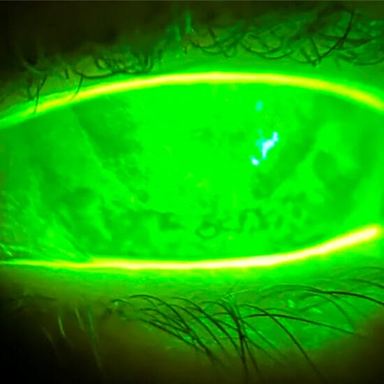
As you can see from the series of photos, there was obvious swelling and hyperaemia of the right upper eyelid and a mild subconjunctival haemorrhage temporally. The anterior eye was otherwise clear. Intra-ocular pressures were R & L 15mmHg. Mydriatic fundus examination revealed no abnormalities.
A black eye, or periorbital haematoma, is the accumulation of blood in the tissue around, but not within the eye
Black Eyes
A black eye, or periorbital haematoma, is the accumulation of blood in the tissue around, but not within the eye. Blunt trauma to the area around the eye can damage the small blood vessels beneath the skin causing them to bleed, leading to oedema and discolouration around the eye.
The periorbital skin is very loose so fluid accumulates easily beneath it. The skin is also very thin in this area, so even a slight pooling of blood can result in significant discolouration. For these reasons, black eyes often appear much worse than they are, as it was in my case. For those of you who hadn’t realised who the girl in the pictures is, the ‘patient’ is in fact me, Jess Chi. The blow of the softball to my brow bone was not of much force, however the oedema and discolouration around the eye following was quite remarkable, which was evident by the reaction of my team.
At the time of injury, ice is the best treatment. Cold therapy to the area reduces blood flow through the ruptured vessels, reducing swelling and the amount of discolouration. Fortunately one of my team members had ice packs to keep the beers cold so I iced the eye while I wasn’t on the field.
Despite its misnomer, a ‘black eye’ usually has little or no effect on the eye itself. Injury to the globe by a blunt object larger than the orbital opening may be absorbed by the orbital contents. Hence, blunt trauma to the orbit may cause damage to the orbit but can spare the eye as it is protected within the orbital cavity. However, blunt trauma can lead to ocular complications, particularly if the object is smaller than the orbital opening, which is why sports such as squash can be more devastating to the eye as its smaller ball size fits well into the anterior part of the orbit.2 Ocular complications can include orbital blow-out fracture, corneal abrasion, lid lacerations, traumatic iritis, anterior hyphaema, traumatic mydriasis, lens subluxation or dislocation, vitreous haemorrhage, retinal detachment and ruptured globe.
Examination
Corrected visual acuity can indicate prognosis and also be used to monitor for progression of damage or secondary complications. If the vision is less than 6/6, pinhole acuity should be performed.
Ocular motility
Reduced eye movements could suggest a ruptured globe, orbital wall fracture (blowout fracture), nerve palsy or retrobulbar haematoma. Blowout fractures will often exhibit enophthalmos. If mobility is restricted, the patient should be referred for further testing, including X-ray or CT scans.
Pupil reactions
Relatively afferent papillary defects could indicate a retinal detachment, vitreous haemorrhage or retrobulbar haemorrhage. Pupil size relative to the uninjured eye should be noted. Traumatic miosis or mydriasis can result, and may be transient, however in cases where the papillae sphincter muscle is damaged, traumatic mydriasis may be permanent and the pupil may react sluggishly or not react at all to light or accommodation.
Anterior Eye examination
Careful slit lamp examination is important to ensure each of the structures of the eye can be visualised and are clear.
Hyphaema can result if damage occurs to the anterior uvea, causing it to bleed. Most will be innocuous and will resolve spontaneously within days, however these should be monitored closely as can lead to secondary haemorrhage or uveitis. Iridodialysis – which is dehiscence of the iris from the ciliary body – can lead to secondary pupils causing monocular diplopia and photophobia.
The lens may be damaged, causing cataract, or damage may occur to the zonules leading to lens subluxation or dislocation. If visually significant, surgery may be required to remove the lens.
Intraocular pressures (IOPs) should be measured and compared to the uninjured eye. Damage to the ciliary body can cause temporary reduction of aqueous secretion causing IOP to decrease, or can lead to anterior chamber angle recession causing IOP to increase.
Fundus Examination
While most of the force of a blunt injury is usually absorbed anteriorly, damage can also occur in the posterior pole. Complications can include posterior vitreous detachment, vitreous haemorrhage, choroidal rupture, retinal detachment, commotio retinae and optic neuropathy. Hence it is important to do confrontational fields and to dilate these patients to ensure no posterior complications have resulted.
Commotio retinae, as a result of contre-coup injury to the eye, usually occurs peripherally, but can affect the macula (causing a cherry red spot). Mild cases usually self-resolve, however more severe cases can lead to
intra-retinal haemorrhage, progressive pigmentary degeneration and macula hole. Optic neuropathy is uncommon but often a devastating cause of permanent visual loss – impact transmit shock wave to the optic canal, damaging the optic nerve. Being relatively myopic, I had concerns of retinal detachment but mydriatic fundus examination ruled this out.
Fortunately, my brow bone and eyelid absorbed all the force from the softball and I had no ocular complications aside from a small subconjunctival haemorrhage which resolved in a few days. My ‘black eye’ lasted about 10 days, and is now fully recovered. So for now, I’ll continue to avoid squash and look to keep my eye on the ball (but not literally).
|
Corrected VA & Pinhole if <6/6 Pupil reactions External examination Eye movements Confrontational fields Mydriatic Fundus examination |
Jessica Chi is the director of Eyetech Optometrists, an independent speciality contact lens practice in Melbourne. She is the current Victorian and National President of the Cornea and Contact Lens Society, and an invited speaker at meetings throughout Australia and beyond. She is a clinical supervisor at the University of Melbourne, and has served on the continuing education committee for the Australian College of Optometry and the Therapeutics Advisory Board for the Optometry Association of Australia
References
1. McCarty CA, Fu CL, et al. Epidemiology of Ocular Trauma in Australia. Ophthalmology 1999; 106:1847-52.
2. Barrell, GV, Cooper PJ et al. Squash ball to eye ball: the likelihood of squash players incurring an eye injury.
Br Med J (Clin Res ED) 1981 Oct 3;283(6296):893–-895 Ehlers JP, Shah CP, Fenton GL, and Hoskins EN. The Wills Eye Manual: Office and Emergency Room Diagnosis and Treatment of Eye Disease (5th edition). Lippincott Williams & Wilkins, 2008.