A well-fitting, well matched ocular prosthesis can dramatically aid the psychological adaptation for a patient who has lost an eye, but patient management is essential.
A consultation with RW, a 75-year-old retired gentleman who wears an ocular prosthesis, provided a timely reminder of the importance of regular examination for prosthetic patients with close examination of not just the healthy eye, but also the prosthetic eye.
RW had been fitted with an ocular prosthesis following enucleation of his left eye for a choroidal melanoma 14 years prior.
He presented for examination, reporting excellent vision with his bifocal spectacles, and no problems with his right eye, however he was finding mucous discharge from his left side. This was worse in the morning but could persist throughout the day.
Most patients with prosthetic eyes will complain of watering, discomfort, discharge and crusting
Upon further questioning, his present ocular prosthesis was 10 years old, and it had been 18 months since his last visit to his ocularist for a polish. RW removes the prosthesis every few months for a clean with Optifree PureMoist contact lens solution.
Examination
RW could achieve spectacle acuity of R 6/5 and N4 at near with his bifocal spectacles of prescription R +1.00/-2.75×99, near addition +2.50DS. No change in his prescription was noted.
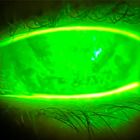
Examination revealed a roughened and deposited surface on the left prosthetic (figure 1), more obvious with sodium fluorescein (Figure 2). Lid eversion revealed giant papillary conjunctivitis in the left eye (Figure 3).
RW was prescribed pred forte to be used twice daily, and it was recommended that he return to his ocularist for a new prosthesis.
Ocular Prostheses
Ocular prostheses are commonly made from polymethyl methacrylate (PMMA). PMMA is chosen because it is inert, biocompatible and thus well tolerated and has low extrusion risk. In addition, it is durable, readily available and relatively inexpensive.
The ocular prosthesis is a ‘shell’ made by impression molding of the socket. The iris is hand painted and the sclera tinted to match the other eye. In the hands of a skilled ocularist, this can give rise to a remarkably close match to the remaining eye, as was the case for RW (Figure 4).
Managing a Prosthetic Eye
Most patients with prosthetic eyes will complain of watering, discomfort, discharge and crusting. One study found that 93 per cent of prosthetic wearers are symptomatic and 60 per cent will experience symptoms daily.1 This is due to the compromised tear films of these patients.
Anopthalmic eyes exhibit less tear production due to absence of the corneal reflex, which is normally responsible for producing much of the aqueous components of tears.2 With reduced aqueous, there will be relatively excessive amounts of mucin and oil components, causing thick mucoid secretions. The aqueous is also important in flushing away debris, hence reduced aqueous will result in greater deposition.
Ocular prostheses can also lead to meibomian gland dysfunction and blepharitis. This is because prostheses cause mechanical trauma to the tarsal conjunctiva, altering the meibomian gland structure. Discharge can also cause meibomian gland obstruction resulting in meibomian gland obstruction.1
Patients may also experience a hypersensitivity response to the prosthesis or to the deposition on the prosthesis, which can result in papillary conjunctivitis. This will lead to irritation and more mucous production.
Treatment for patients experiencing symptoms of dryness, irritation and discharge depends on the severity. In mild cases, viscous ocular lubricants may suffice. In cases of allergic conjunctivitis, topical anti-histamine and mast-cell stabiliser combinations such as Patanol or Zaditen may be useful, but more pronounced cases may warrant topical NSAIDs or topical steroids. Topical steroids have frequent side effects including ocular hypertension and cataract formation, however this is not an issue for patients with ocular prostheses as they are worn for anophthalmic or disfigured non-seeing eyes.
It is also important to ascertain how often the prosthetic is removed and how it is cleaned. This will vary depending on the individual. Patients wearing a prosthesis on an existing eye will experience more awareness than an anophthalmic patient, so will require removal and cleaning more frequently – often daily. If the patient is anophthalmic and maintains good quality tears, the prosthetic only needs to be cleaned every one to two months.
Tear deficiency and allergy usually results in a stringy white discharge. However, green or yellow discharge accompanied with hyperaemic orbital tissue can indicate infection, and topical antibiotics should be prescribed.
Discomfort may also arise due to problems with the orbital area, including malignancy, systemic disease, contracted socket syndrome and implant exposure. Any suspicion of malignancy, particularly in patients who have had a history of malignancy should be referred immediately to their ophthalmologist. The orbital structure may change with time and may contract or adhesions can form within the tissue. These patients may require surgical intervention or a new ocular prosthesis.
Care and maintenance
Cleaning should be performed by rubbing gently with a soft multipurpose solution (MPS), wiping with a wet tissue, and then rinsing with fresh MPS. If left out overnight the prosthesis should be stored in MPS. If a patient is sensitive to preservatives, the prosthesis should be rinsed with saline prior to insertion.
It is recommended that a patient sees their ocularist at least annually for polishing and adjustments, with replacement approximately every five years to accommodate for changes to the eye socket.3
Ocular Melanoma
Choroidal melanoma is the most common primary malignant intraocular tumour, with the incidence of ocular melanoma in Australia reported at eight per million men and six per million women. The incidence is highest in males aged over 65, with a greater incidence in rural areas compared to urban. The incidence is also higher in Caucasian individuals, patients with light coloured irises, and patients that have had higher amounts of UV exposure.4
Choroidal melanoma has serious implications, with a mortality rate of 30–50 per cent within 10 years of diagnosis.5 Death is usually a result of distant metastases, hence early detection and prompt treatment is crucial. Treatment for choroidal melanoma is to remove the melanoma, either via enucleation or globe-sparing treatments for smaller tumours such as brachytherapy or radiation. Unfortunately, choroidal melanomas often metastasise prior to diagnosis. Australia suffers the highest rate of melanoma in the world, hence the importance of UV protection and regular exam.
Fortunately for RW, his melanoma was detected prior to any metastases.
Psychological Adaptation
Loss of an eye can be extremely challenging for a patient psychologically and emotionally. Having a well-fitting ocular prosthesis that is well matched to the remaining eye can dramatically aid the psychological adaptation for the patient. Fourteen years later, RW is retired, fully adjusted to being monocular and reports clear comfortable vision. Most people he meets are completely unaware he is wearing an artificial eye, largely due to the expert work of his ocularist Mr. Patrick Loyer.
Jessica Chi is the director of Eyetech Optometrists, an independent specialty contact lens practice in Melbourne. She is the current Victorian and National President of the Cornea and Contact Lens Society, and an invited speaker at meetings throughout Australia and beyond. She is a clinical supervisor at the University of Melbourne, and has served on the continuing education committee for the Australian College of Optometry and the Therapeutics Advisory Board for the Optometry Association of Australia
References
1. Pine K, Sloan B, Stewart J, Jacobs RJ. Concerns of anophthalmic wearing Artifical Eyes. Clin Exp Ophthal 2011; 39: 47-52
2. Allen et al. Artificial eyes and tear measurements. Ophthalmology 1980; 87:155-157
3. Cafiero-Chen M et al. Ocular Prosthesis: Indications to Management. Canadian Journal of Optometry Vol. 77 issue 2
4. Vajdic CM, Kricker A, Giblin M, McKenzie J, Aitken J, Giles GG, Armstrong BK. Incidence of ocular melanoma in Australia from 1990 to 1998. Int J Cancer. 2003;105:117–22.
5.Ten-year follow-up of fellow eyes of patients enrolled in Collaborative Ocular Melanoma Study randomised trials: COMS report no. 22. Ophthalmology. 2004 May. 111(5):966-76.