In my October column I wrote about Jason, who has been managed in my practice for 26 years. Now we see his two sons… demonstrating the rewards that come from building strong relationships.
Harry first presented to my practice aged 16. He’d come in because I’d suggested to his father, Jason, that his sons should be examined. Jason has keratoconus and I was concerned that they might develop the same condition.
Harry had never had his eyes examined and he reported no problems with his vision, although he experienced itchy eyes, and admitted to eye rubbing. He suffers from hayfever, but is otherwise in good health.
Vision unaided was R 6/6+ and L 6/7.5+.
Subjective refraction gave
R +0.25/-0.50×165 (6/4.8) and
L +0.25/-0.75×158 (6/6=).
Relatives of keratoconus patients are 15–67 times more likely to develop the condition
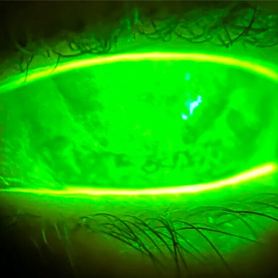
Anterior eye examination revealed clear corneas in both eyes, with no ocular surface staining. The tear film was unstable with a tear film break up time of four seconds in both eyes. Lid eversion revealed grade two papillary conjunctivitis in both eyes (Figure 1).
Corneal topography with the Medmont E300 topographer revealed a mild sagging cone in Harry’s left eye and forme fruste in the right eye (Figure 2).
I prescribed him Patanol twice daily to control his ocular allergies, and counselled him about the importance of avoiding eye rubbing. A review was scheduled for three months.
Harry returned as scheduled, reporting no problems with his vision. He had been consciously trying to avoid eye rubbing but found himself doing so, on occasion, out of habit.
Vision unaided was unchanged at R 6/6+ and 6/7.5+.
Subjective refraction revealed a mild increase in Harry’s left astigmatism to L +0.25/-1.00×162 (6/6=). The right was unchanged.
Corneal topography revealed keratoconic progression in the left eye, with the difference map from this exam to the prior exam revealing steepening of Harry’s left cone by four dioptres (Figure 3).
Harry was counselled again not to rub his eyes, and referred to a corneal specialist for corneal crosslinking (CXL), which was performed one month later.
DISCUSSION
Keratoconus is a non-inflammatory, progressive condition, which is normally bilateral but asymmetric, and typically manifests in the late teens to early adulthood.1 The aetiology is uncertain, however it is considered to be multifactorial – with genetic, biochemical, environmental, and behavioural factors. It has been reported that six to 23.5 per cent of patients with keratoconus have a positive family history. Relatives of keratoconus patients are 15–67 times more likely to develop the condition.2,3
Extensive research into the genetics of keratoconus has implicated various genes and inheritance patterns however inheritance patterns vary between families and is unfound in others.2,3
Progression
Keratoconus tends to progress fastest in the younger years, then stabilising by the late to mid-30s.5 This is a result of the cornea stiffening with time as natural crosslinks form between the collagen fibrils in the cornea.
In the early stages of keratoconus, patients may be asymptomatic. However, as the disease becomes more pronounced and corneal irregularity increases, patients may experience symptoms of distorted vision, glare sensitivity, ghosting, shadowing, and haloes, along with generally reduced quality of life. Spectacles may assist in the early stages, but as the condition progresses, rigid contact lenses may be required to correct vision.
Because keratoconus tends to progress more quickly in the younger years, young patients must be monitored closely.5 Fortunately, with technology such as corneal topographers, the condition may be detected in the early stages, and CXL can be done to intervene so that patients may be spared visual loss.
Corneal Crosslinking
CXL with riboflavin and UVA has been shown to be effective in halting progression of keratoconus and can even result in some regression. CXL increases the rigidity of the cornea, accelerating the natural crosslinking that occurs with time.6,7 The current standard protocol for CXL involves removing the corneal epithelium prior to treatment to ensure the riboflavin penetrates into the corneal stroma. Following, many patients experience pain until the cornea re-epithelises, with the level of pain varying from mild to extreme. While CXL has been shown to be effective at halting the progression of keratoconus, it is not without risk of complications. Following the procedure, there will be corneal stromal haze, which may be permanent in some. There have also been reported cases of microbial keratitis and other complications. Hence, CXL should be reserved for progressive cases of keratoconus.
Reduce Risks
While there are still many unknowns about the exact aeitology of keratoconus, we do know that relatives of patients with keratoconus are much more likely to suffer from the condition. For this reason, family members should be screened, especially in the teenage years when the condition tends to manifest and is more rapidly progressive.
Ocular allergies and eye rubbing have been associated with the pathogenesis of keratoconus,4 and as such, patients must be counselled about the need to avoid eye rubbing. Every keratoconic patient in my practice is asked whether they rub their eyes, and they, along with their family members, are advised to avoid doing so, as this is the only patient modifiable risk factor that can prevent progression. If ocular allergies are present, therapeutic agents such as Patanol – a combination of an anti-histamine and mast cell stabiliser – can be prescribed to reduce the symptoms and help to reduce the temptation to rub eyes. Oral anti-histamines and cold compresses may also alleviate symptoms. In more severe cases, topical steroids can be administered.
BACK TO HARRY… AND ALBERT
Luckily for Harry, early detection of keratoconus and intervention spared him any requirement for visual correction.
I also examined Harry’s older brother Albert (aged 20), who fortunately exhibited no signs of any keratoconus. Now my aim is to review Albert on an annual basis in case he goes on to develop the condition.
Jessica Chi is the director of Eyetech Optometrists, an independent specialty contact lens practice in Melbourne. She is the current Victorian, and a past National President of the Cornea and Contact Lens Society, and an invited speaker at meetings throughout Australia and beyond. She is a clinical supervisor at the University of Melbourne, a member of the Optometry Victoria Optometric Sector Advisory Group and a fellow of the Australian College of Optometry and the British Contact Lens Association. Jessica writes ‘mipatient’ on alternate months with Margaret Lam.
References
- Rabinowitz YS. Keratoconus. Surv Ophthalmol 1998;42:297-319.
- Rabinowitz YS. The genetics of keratoconus. Ophthalmol Clin North Am 2003 16:607-620
- Wheeler J, et al. The Genetics of Keratoconus: A Review. Reprod Syst Sex Disord. 2012 Jun 3;(6)pii:001
- McMonnies CW, Boneham GC. Keratoconus, allergy, itch, eye-rubbing and hand-dominance. Clin Exp Optom 2003;86(6):376-384.
- Tuft SJ, Moodaley LC, Gregory WM, Davison CR, Buckley RJ. Prognostic factors for the progression of keratoconus. Ophthalmology. 1994;101(3):439–447.
- Witting S, et al. A randomized controlled trial of corneal collagen cross-linking in progressive keratoconus: preliminary results. J Refract Surg. 2008 Sep;24(7):S720-5.
- Meek K, Hayes S. Corneal cross-linking – a review. Ophthalmic Physiol Opt 2013,33,78-93.