Vitreoretinal surgeon and ophthalmologist Professor Andrew Chang AM (above) discusses how, for many years, the Optos ultra-widefield technology has helped him diagnose, monitor, and treat retinal disease.
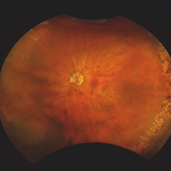
As rates of diabetes continue to rise globally, the early detection and management of diabetic eye disease has become an increasingly important focus for eye care professionals. Retinal imaging technologies, such as Optos ultra-widefield (UWF) imaging, are changing the way clinicians document and assess the retina, particularly by capturing up to 200° of the retina in a single image.
Prof Andrew Chang said UWF imaging enables clinicians to capture a much broader view of the retina than traditional imaging methods, while also creating the digital platform for monitoring disease progression over time.
“Ultra-widefield digital imaging has revolutionised how ophthalmologists and eye care professionals work,” he said. “In my practice, I have used the Optos since its arrival in Australia, to image the retina in a much wider field. It is an accessible, efficient way to assess patients in the clinic.”
In diabetic retinopathy, the initial challenge is to make the diagnosis and detect the degree of diabetic eye disease in a screening capacity
Improving Diabetic Retinopathy Assessment
Diabetic retinopathy presents several challenges, Prof Chang explained, beginning with screening and diagnosis before moving into the assessment of retinal damage and circulation.
“In diabetic retinopathy, the initial challenge is to make the diagnosis and detect the degree of diabetic eye disease in a screening capacity,” Prof Chang said. “Then we move next to imaging and evaluating the vascular circulation and ischaemia of the neural retina by performing fluorescein angiography.”
Optos said its devices are designed to support ocular assessment by providing high-resolution UWF retinal images in a single shot. The company said optomap can capture 82% of the retina with a 200° image, in a single capture in each imaging modality.
Prof Chang said the imaging system is also capable of performing fluorescein angiography, allowing ophthalmologists to study the circulation in greater detail of the peripheral retina.
Capturing the Retinal Periphery
Follow-up monitoring remains a critical component of diabetes-related eye care, particularly identifying disease progression and determining when intervention is required. Prof Chang said clinicians strive to screen for the development of retinopathy, follow any progression in severity, and grade disease using diabetic retinopathy severity scores.
He said imaging technology has improved the ability to document diabetic retinopathy compared with older methods.
“The traditional way that we diagnose or record diabetic retinopathy is a seven-field montage,” he said. “Now with a single image, Optos lets us capture those seven fields and more.”
According to Prof Chang, the ability to visualise the retinal periphery has become increasingly important, particularly as clinicians learn more about the relationship between peripheral vitreoretinal disease and progression of proliferative retinopathy. Missing these peripheral lesions results in blindness from untreated progression of retinopathy.
For patients that have already developed retinopathy, assessment of the periphery is important because we can potentially treat areas of non-perfusion earlier, with targeted laser treatment
Earlier Intervention and Treatment
Assessment of retinal perfusion is another important factor in diabetic eye disease management. Prof Chang explained that colour photography can show haemorrhages and imply perfusion, while fluorescein angiography allows clinicians to study retinal circulation in real time, showing vascular wall damage and leakage, and ischaemia.
“The assessment of the peripheral perfusion is important,” he said. “When we are studying the circulation of the retina, which is what’s affected in diabetic retinopathy, we have detailed information from fluorescein angiography.”
Prof Chang said approaches to diabetic retinopathy treatment are continuing to evolve, with current clinical trials investigating earlier intervention strategies aimed at slowing disease progression.
“We are now recruiting patients into trials where we’re looking at different ways of treating diabetic retinopathy earlier, in the hopes that it will retard the progression and blindness,” he said.
He added that identifying areas of retinal non-perfusion can help guide targeted treatment.
“For patients that have already developed retinopathy, assessment of the periphery is important because we can potentially treat areas of non-perfusion earlier, with targeted laser treatment,” he explained.
Enhancing Clinical Workflow and Patient Care
Prof Chang described the benefits of Optos UWF imaging in the clinic as “multifaceted”, citing efficiency, patient comfort, and improved workflow.
“The first (benefit) is we have gathered information about a patient progressively during the course of their disease very efficiently for diagnosis and follow up, but for a patient also, it’s a comfortable way of having their eye examined, even through an undilated pupil,” he said. “For a clinic, the efficiency of moving a patient in and out through a designated screening area or in a separate imaging area is also excellent.”
Optos described its imaging as fast and patient friendly, with some devices producing a 200° single-shot optomap retinal image in less than half a second.
Prof Chang said digital imaging also plays an important role in patient education and collaboration between healthcare professionals.
“The virtual digital platform that Optos allows is really valuable to show patients to assist in understanding their disease,” Prof Chang said. “The other major advantage is that we can share information digitally, which allows eye care professionals to collaborate and synergise care.”
While advances in imaging and treatment continue, Prof Chang said one of the greatest ongoing challenges is ensuring patients are referred early for diagnosis and intervention.
“We still unfortunately, see patients present with advanced blinding and preventable diabetic eye disease,” he said.
Despite this, he remains optimistic about the impact of earlier diagnosis and treatment.
“Globally, diabetic retinopathy is a blinding disease, but it is a treatable disease and if we diagnose early, we can treat early and effectively, and we can prevent patients from losing sight.”