

Epiretinal membrane is one of the commonest retinal diseases and a frequently under-recognised cause of impaired vision in clinical practice.2-5 Management ranges from simple observation through to surgery.
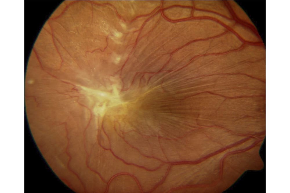
Epiretinal membranes (ERMs) are fibrotic sheet-like membranes that develop on the macula surface (Figure 1). They may cause decreased visual acuity (VA) and metamorphopsia, potentially leading to decreased quality of life.1
Posterior vitreous detachment (PVD) is present in 70% to 95% of patients with idiopathic ERM …

Figure 1. Epiretinal membrane.
ERMs have been called a variety of names including cellophane maculopathy, pre-macular fibrosis, epiretinal fibrosis, epimacular membrane, surface wrinkling retinopathy and macular pucker.6 These terms reflect the clinicopathologic findings produced by ERMs of varying severity.
They are classified as either ‘idiopathic’ or ‘secondary’ to another ocular condition.
EPIDEMIOLOGY
The Australian Blue Mountains Eye Study reported a 7% prevalence of ERM in people aged 49 years or older.7 Age is a significant risk factor for ERM with the prevalence peaking at 12% between the ages of 70–79 years, 31% of which are bilateral.5,8-11 Both sexes appear to be affected equally.
Posterior vitreous detachment (PVD) is present in 70% to 95% of patients with idiopathic ERM and may play an important role in its development. This correlates with the higher prevalence of ERM in older patients as they are more likely to have developed a PVD than younger patients.7,12 Correspondingly, ERMs are uncommon in children and young adults in the absence of predisposing conditions such as uveitis or trauma.

Figure 2a. Before PVD onset showing no epiretinal membrane.
ERMs have been reported to develop in 11% of patients by three years following cataract surgery.9 The acceleration of PVD formation by cataract surgery is considered the most likely contributing factor.
PATHOGENESIS
ERMs result from fibroglial cellular proliferation on the retinal surface. They have contractile properties that can lead to distortion and thickening of the retina, resulting in blurred vision or metamorphopsia.
Our understanding of the pathogenesis of idiopathic ERM is evolving. ERM development has long been linked to the presence of a PVD. According to the classic hypothesis, tractional forces at the vitreoretinal interface during a PVD cause breaks in the internal limiting membrane (ILM) through which glial cells from the inner layers of the retina migrate onto the retinal surface.13 The proliferation of these cells then leads to the formation of an ERM.

Figure 2b. One week after PVD onset. Early epiretinal membrane is present.
CASE STUDY ONE: ERM ASSOCIATED WITH PVD
A 61 year old woman reported a ten month history of mild left visual distortion. Twelve months previously, her optometrist had diagnosed a left PVD after she had complained of left flashing lights and floaters.
MANAGEMENT
Examination demonstrated a left VA of 6/5 associated with a PVD and ERM with a macular pseudohole. No other retinal pathology was detected. Due to minimal symptomatology and excellent VA, observation was recommended.
OUTCOME
Two years later the VA remained 6/5 with no subjective change in symptoms.
Serial retinal photos taken before the onset of PVD, one week following PVD symptoms developed and two years later, show the initial development of a mild ERM with cellophane appearance and its subsequent progression to a macular pseudohole (Figure 2a, b, c).

Figure 2c. Two years after PVD onset. Established epiretinal membrane with pseudohole has developed.
DISCUSSION
PVD is present in the majority of patients with idiopathic ERM and is believed to play an important role in ERM development. Patients with excellent VA and minimal symptoms can be monitored without treatment.
Specimens obtained during vitrectomy surgery show that ERMs consist of glial cells, retinal pigment epithelial cells, macrophages, fibrocytes, and collagen. The relative proportions of these cells vary according to the aetiology of the ERM. Those associated with retinal breaks or retinal detachment (RD) are composed mainly of dispersed RPE cells, while cells of glial origin predominate in idiopathic ERM.
A more recent alternative hypothesis suggests that remnants of the posterior vitreous cortex may remain attached to the ILM after PVD.14 Mechanical traction induced by the PVD is believed to stimulate the production of growth factors which activate hyalocytes within the cortical vitreous remnants. This causes the hyalocytes to proliferate and differentiate into myofibroblasts, leading to ERM formation.
Inflammatory mediators produced through concurrent intraocular disease may also promote fibrocellular growth, leading to secondary ERM formation. Secondary ERMs are associated with a variety of ocular conditions including retinal tears, RD (clinically significant ERMs develop in up to 9% of eyes after RD surgery), vitreous haemorrhage, retinal vascular occlusive diseases (central and branch retinal vein occlusion), diabetic retinopathy, ocular inflammatory diseases, retinitis pigmentosa, ocular trauma, intraocular surgery and intraocular tumours (Table 1).

Figure 3a. Epiretinal membrane with collateral vessels below the optic disc.
CASE STUDY TWO: ERM SECONDARY TO A BRANCH RETINAL VEIN OCCLUSION
A 70 year old woman was referred with a 12 month history of progressively blurred left vision and distortion. She had a previous history of a left inferotemporal branch retinal vein occlusion which developed five years prior and had resolved without treatment. She was on treatment for hypertension and hypercholesterolemia.
MANAGEMENT
Examination demonstrated a left VA of counting fingers only, associated with an advanced ERM (Figure 3a). Collateral retinal vessels were visible inferior to the optic disc and consistent with a history of previous branch retinal vein occlusion. A 25 gauge vitrectomy with peeling of the ERM and ILM was performed.

Figure 3b. Three months after epiretinal
membrane surgery.
OUTCOME
At three months post-op, the left VA had improved to 6/12 and the patient reported a significant reduction in distortion (Figure 3b).
DISCUSSION
Retinal vascular diseases such as branch retinal vein occlusion, central retinal vein occlusion and diabetic retinopathy are frequently associated with chronic retinal vascular leakage, which can lead to development of a visually significant secondary ERM.
PROGNOSIS
The effect that an ERM has on vision is highly variable and determined by the severity of the distortion that it causes, the location of the ERM in relation to the fovea, the thickness of the ERM, and the presence or absence of macular oedema.

Table 1
ERM is a chronic condition with a typically slow onset and progression. Many patients stabilise with time, and regression of clinical signs can occur.
CASE STUDY THREE: ERM WITH SPONTANEOUS AUTO-PEELING
A 45 year old woman was referred with a three month history of slightly blurred right vision and distortion.
MANAGEMENT
Examination showed a right VA of 6/7.5 and an ERM (Figure 4a). No other retinal pathology was visible. As the patient reported only mild symptoms with excellent VA, a period of observation was recommended to detect possible progression of the ERM.
OUTCOME
The patient was reviewed at six monthly intervals. Over a period of two years, the ERM underwent spontaneous release due to autopeeling. The VA improved to 6/6. (Figure 4b)

Figure 4a. Epiretinal membrane.
DISCUSSION
Spontaneous auto-peeling and ERM release can occur in approximately 2% of patients. The mechanism may involve contraction of the ERM without PVD in some patients and development of a PVD in others. Despite auto-peeling occurring, recurrent ERMs can still develop in these eyes so continued monitoring is recommended.
The Blue Mountains Eye Study found that over a five year period, approximately 9% of patients with mild ERMs without retinal folds, progressed to more severe ERMs associated with retinal folds and 13.5% of patients with an ERM in one eye, developed an ERM in the fellow eye.15
The average reduction of VA over five years was 2.8 letters in eyes with mild ERMs without retinal folds and 7.4 letters in eyes with ERMs and retinal folds. A reduction of VA by five or more letters (one line or greater) was observed in 43% of eyes with ERM and retinal folds.

Figure 4b. Two years later showing spontaneous
auto-peeling of the epiretinal membrane.
CLINICAL PRESENTATION
History
Patients typically present with a gradual onset of metamorphopsia or blurring of vision which is usually mild but can sometimes be severe. In the early stages, ERMs are often asymptomatic and may be detected during a routine eye exam, particularly when optical coherence tomography (OCT) scanning is performed.
Although the majority of ERMs are idiopathic, it is important to take a targeted history to identify conditions that can cause secondary ERMs (Table 1). Patients should be asked about flashing lights or floaters (associated with PVD, retinal tear or RD), any history of ocular trauma, intraocular surgery (particularly cataract surgery), retinal vascular disease or uveitis.
With progression and contraction of the ERM, patients may notice increasing metamorphopsia. Aniseikonia or blurred vision may be noted. Central photopsia and monocular diplopia are infrequently reported.

Figure 5. Grade 0 epiretinal membrane (cellophane
maculopathy).
Examination Findings
The clinical signs of an early ERM can be subtle and difficult to detect on fundus examination but become more obvious as the ERM progresses and thickens.
A three step grading system proposed by Gass remains the standard for classification of ERM’s.16 Although it is not typically used in clinical practice, it provides a useful overview of clinical signs to look for when assessing ERM’s (Table 2).
Grade 0 (cellophane maculopathy) is characterised by an early ERM identified by the presence of a fine, transparent membrane over the inner retinal layer, which looks similar to a piece of cellophane. As there is no retinal distortion, the patient is usually asymptomatic (Figure 5).

Figure 6. Grade 1 epiretinal membrane (crinkled
cellophane maculopathy).
Grade 1 (crinkled cellophane maculopathy) is associated with ERM contraction causing fine superficial retinal folds that radiate out from the margins of the ERM leading to an irregular wrinkled appearance (Figure 6). The patient may notice visual changes at this point as ERM contraction distorts the distribution of photoreceptors in the fovea. This can affect image perception causing objects to appear distorted (metamorphopsia), larger (macropsia) or smaller (micropsia). Some of the symptoms associated with ERM may result from aniseikonia (Greek: anisos, unequal; eikon, image) or the perception of the same image as being of different sizes with each eye. Note, in unilateral cases, patients with advanced ERM development may not necessarily report symptoms, especially if the non-dominant eye is affected.
Grade 2 (preretinal macular fibrosis or macular pucker) is used to describe a thicker, more opaque ERM, which causes full thickness retinal distortion and obscures the underlying retinal vessels which also appear increasingly tortuous (Figure 7). Over three quarters of patients with grade 2 ERM experience symptoms of blurred vision or distortion.17
An Amsler grid is used to identify and document any visual distortion. Slit-lamp examination should be performed to identify signs of uveitis (e.g. anterior chamber or vitreous cells, keratic precipitates or posterior synechiae). The anterior vitreous should be examined to exclude the presence of brown pigmented cells (often called tobacco dust) which if present, is known as a positive Shafer’s sign (Figure 8). The pigmented cells represent retinal pigment epithelial cells that have passed through a retinal tear into the vitreous cavity. When a positive Shafer’s sign is present, a peripheral retinal tear can be identified in over 90% of patients.

Figure 7. Grade 2 epiretinal membrane (macular pucker).
It is important to examine the peripheral retina to exclude retinal tears in all ERM patients because retinal tears are the commonest cause of secondary ERM. This is ideally performed using a binocular indirect ophthalmoscope with scleral indentation.
CASE STUDY FOUR: ERM ASSOCIATED WITH PERIPHERAL RETINAL TEAR
A 55 year old was referred with a six month history of mildly blurred right vision.
He reported no other symptoms.
MANAGEMENT
Examination of the right eye demonstrated a VA of 6/7.5. Amsler grid testing showed mild distortion. A PVD with vitreous tobacco dusting (positive Shafer’s sign) and an ERM were present. Peripheral retinal examination revealed a superotemporal horseshoe retinal tear with a surrounding cuff of subretinal fluid. (Figure 10a).
The retinal tear was treated with laser to prevent retinal detachment (Figure 10b).
The ERM was monitored for progression.

Figure 8. Vitreous tobacco dust.
OUTCOME
The patient’s clinical condition remained stable over the following two years with no change in symptoms, VA or clinical appearance of the ERM.
DISCUSSION
This case highlights the importance of examining the peripheral retina to exclude the presence of a retinal tear in all patients with an ERM. Retinal tears often lead to dispersion of retinal pigment cells into the vitreous cavity, which in turn can lead to ERM formation.
Macular pseudoholes are discrete round reddish lesions in the central macula commonly seen with ERMs. They are caused by centripetal contraction of the ERM with slight central movement of the underlying retina and steepening of the foveal margin, which creates the appearance of a hole.18 They look similar to full thickness macular holes on slit lamp examination (Figure 9a & 9b).
It is important to distinguish a macular pseudohole from a true full thickness macular hole because the latter generally leads to progressive loss of vision and benefits from prompt surgery, whereas macular pseudoholes have a better visual prognosis and are less likely to require surgery. Macular holes are full thickness defects in the macula, caused by vitreomacular traction.

Table 2
The Watzke-Allen test is a simple, effective clinical test to differentiate between a macular pseudohole or macular hole, and is particularly useful when OCT is unavailable. The test is performed at the slit-lamp by visualising the macula with a fundus lens and projecting a thin bright slit beam (vertically and then horizontally) across the suspected macular pseudohole or macular hole. Patients with macular pseudoholes describe the slit beam as looking straight and complete because retinal photoreceptors are present in the base of the pseudohole and capture the beam. In contrast, patients with a macular hole report seeing a broken or kinked slit beam because photoreceptors are absent at the base of a macular hole (Figure 11), (Table 3).

Figure 9a. Macular pseudohole.
Retinal Imaging
Fundus photography can aid diagnosis of an ERM, help to document disease progression, and assist in patient education. Red free imaging may improve visibility of ERMs and highlight retinal vascular changes (Figure 12a & 12b).
Fluorescein angiography (FA) may be performed if underlying conditions are suspected to contribute to vision loss and ERM formation. These conditions include diabetic retinopathy, cystoid macular oedema and retinal vein occlusions which may be associated with vascular leakage demonstratable with FA (Figure 13a & 13b).6
OCT is very helpful in the diagnosis and monitoring of ERMs and is more sensitive than fundus photography for identification of ERMs. Features of an ERM on high resolution cross sectional macular scans include a hyper-reflective line on the retinal surface, saw-tooth wrinkling of the retinal surface, elevation or loss of the foveal dip and in some cases intraretinal oedema and subretinal fluid (Figure 14). OCT can easily distinguish between a macular pseudohole and a macular hole (Figures 9b & 11).

Figure 9b. OCT scan of a macular pseudohole.
OCT macular thickness maps provide a topographical display of the macular thickness and highlight areas of retinal thickening in areas affected by ERM contraction. They provide objective and quantitative information which enables reliable assessment of disease progression. ERM progression is characterised by increasing macular thickness measurements on serial OCT scans. Following ERM surgery, OCT macular thickness maps can be used to monitor the response to surgery and typically show a progressive reduction in macular thickness measurements during the first postoperative year (Figure 14).

Table 3
MANAGEMENT
Not all ERMs produce visual symptoms or need surgery. Patients with normal VA and minimal symptoms can be monitored by an optometrist every six to 12 months. They should be taught how to use an Amsler grid to monitor their central vision and identify visual distortion. OCT is particularly helpful to objectively monitor the condition.
Development or worsening of symptoms (blurred vision, metamorphopsia or aniseikonia), reduction in VA or progressive macular thickening on serial OCT are indicators of ERM progression and should prompt a discussion regarding the pros and cons of ERM surgery.

Figure 11. OCT scan of a macular hole.
When to Perform ERM Surgery
The standard treatment for visually significant ERM is a pars plana vitrectomy (PPV) with ERM peeling. ERM is the third most common indication for PPV.19
Indications for ERM surgery include a deterioration in VA or increasing symptoms. The decision regarding when to perform surgery should be carefully balanced and consideration should be given to the individual patient’s symptoms, clinical findings, visual needs, level of risk aversion, lifestyle, occupation, and personal preferences.
The decision to recommend surgery is relatively straightforward in eyes with rapidly progressing ERMs causing decreased VA. These eyes usually show substantial VA improvements since macular damage from the epiretinal membrane is short-lived and macular function recovers well.

Figure 12a. Epiretinal membrane.
In the past, vitreoretinal surgeons used an arbitrary VA cut-off such as 6/18 to determine who should be offered ERM surgery. However, improvements in surgical technology, better outcomes, and increasing patient expectations have led to reduced thresholds for ERM surgery. It should be remembered that ERMs may significantly impair quality of vision and quality of life due to interference with reading, working, driving, or other tasks. These symptoms are not adequately measured by VA charts thus VA alone should not be used as a determinant of the need for surgery.
In eyes with VA of 6/9 or better and progressive ERM, should surgery be performed early or delayed until the VA decreases to 6/18? Research confirms that the potential for improvement decreases with prolonged observation and the best predictors of outcome following ERM surgery are preoperative VA and duration of symptoms. If surgery is delayed until VA decreases from 6/9 to 6/18 over a period of five years, the visual result will be less favourable than if surgery was performed when the VA was 6/9.
ERM surgery has been shown to produce a remarkable improvement in patients’ quality of life and earlier surgical intervention has been shown to be associated with better VA outcomes.1,20-25 My decision to offer surgery is based on patient symptoms and visual function. I often perform ERM surgery in patients with good VA with significant distortion or an occupational requirement for excellent VA or stereopsis.

Figure 12b. Epiretinal membrane with red free imaging.
CASE STUDY FIVE: ERM IN A JEWELLER WITH GOOD VA
A 58 year old jeweller complained of increasing difficulty seeing fine details on jewels during the past year.
MANAGEMENT
His VA was 6/6 OD, 6/9 OS. Examination showed an isolated left ERM (Figure 16a). Sutureless vitrectomy with ERM peeling was performed.
OUTCOME
The left VA improved to 6/6 at two months post-op and the patient reported a significant subjective improvement in quality of vision making his work easier (Figure 16b).
DISCUSSION
This case highlights the potential for early ERM surgery to preserve and improve visual function in symptomatic patients with high visual demands and good VA.

Figure 13a. Epiretinal membrane.
Detailed assessment of OCT scans may provide prognostic information as certain OCT features may help predict which patients will have better outcomes after ERM surgery.22-25 The thickness of the outer nuclear layer and ellipsoid band integrity best predicts VA pre- and postoperatively.26 The inner nuclear layer thickness best predicts the degree of metamorphopsia both before and after surgery.27
ERM Surgery
Vitrectomy surgery with ERM peeling is a common and effective treatment for ERM which reduces distortion, avoids the risks of ERM progression and often improves VA. It is a form of keyhole surgery, routinely performed as a day case procedure, and typically using local anaesthesia. Additional intravenous sedation is effective in relaxing even the most anxious patients. The procedure can also be performed under general anaesthesia depending upon patient and surgeon preference.

Figure 13b. Epiretinal membrane with vascular leakage demonstrated on fluorescein angiography.
Sutureless vitrectomy techniques using 23g, 25g or 27g trocar systems are typically employed. Compared to traditional 20g vitrectomy techniques, which require suturing of scleral entry wounds, a sutureless vitrectomy enables a more rapid, comfortable recovery due to the absence of suture related foreign body sensation, and high rates of patient satisfaction. The procedure consists of performing a core vitrectomy and induction of a PVD if one is not already present. The ERM is then removed by peeling it off the retinal surface using microforceps (Figure 17).
ERM surgery is often combined with additional procedures such as the use of retinal dyes to enhance visualisation of the ERM or internal limiting membrane (ILM), peeling of the ILM, and/or cataract surgery. The ILM is the anatomical boundary between the vitreous and the retina. Histological data shows that the ILM acts as a scaffold for cellular proliferation and ERM formation. The benefit of ILM peeling during ERM surgery remains controversial.
The idea behind removing the ILM is based on the finding that following ERM peeling alone, fragments of ERM have been observed in approximately 80% of eyes, indicating that ERM peeling has been incomplete with a risk of ERM recurrence if the residual ERM fragments proliferate over time. By removing the ILM, a complete ERM peel can be achieved and the anatomical scaffold for ERM growth is removed. This reduces the rate of post-operative ERM recurrence, from between 12% to 17% of eyes undergoing ERM removal alone, compared to 2% to 4% with ERM and ILM removal.28

Figure 14. OCT showing an epiretinal membrane.
Studies have provided conflicting results regarding whether ILM peeling impacts visual outcomes, but a systematic review of comparative studies indicated that removing the ILM does not appear to affect VA outcomes.29
The use of surgical dyes to stain the ERM and ILM has been considered controversial due to potential retinotoxicity associated with certain dyes such as indocyanine green (ICG), however newer retinal dyes such as trypan blue and brilliant blue G appear to be safe and potentially reduce iatrogenic retinal trauma by enhancing visualisation of the ERM and ILM during surgery (Figure 18).30
Intraocular air or gas are not typically needed during ERM surgery but may be used to tamponade the retina if a retinal tear or retinal detachment are identified per-operatively. Some surgeons routinely use intraocular air to encourage closure of self-sealing sclera incisions and reduce the risk of post-operative wound leak.
If an air or gas bubble is used, the patient is advised to avoid travelling to altitude or flying for a period of two to six weeks post-operatively (depending upon the type of gas used). Vision is poor while the gas bubble is present but recovers when the bubble has spontaneously resorbed.

Figure 15. OCT macular thickness map showing macular thickening before and after epiretinal membrane peeling surgery.
If the patient has a visually significant cataract, consideration should be given to whether to perform cataract surgery before ERM surgery, in combination with ERM surgery, or after ERM surgery. The benefits of combined cataract and ERM surgery include a reduced number of surgical procedures with fewer total post-operative visits and faster visual recovery. The potential downsides of combined surgery are that it is a more invasive procedure with a greater risk of post-operative uveitis, IOL calculations are marginally less accurate, and it may not be available as many vitreoretinal surgeons do not perform cataract surgery.
Variations between surgeons include the vitrectomy technology used (20g, 23g, 25g, or 27g), whether or not triamcinolone acetonide is used to highlight the vitreous and confirm the presence of a PVD, whether or not retinal dyes are used to visualise the ERM and ILM, the type of dye used (e.g. indocyanine green, trypan blue, Brilliant Blue G), whether or not the ILM is peeled, the extent of ERM and ILM peeling (e.g. limited to one disk diameter around the fovea or a larger area), the use of tamponade agents (air, SF6, C2F6, C3F8) and the use of combined cataract surgery.

Figure 16a Epiretinal membrane before surgery.
Results of ERM Surgery
Patients typically experience a gradual improvement in visual function over the course of the first postoperative year. Metamorphopsia symptoms generally improve but do not resolve completely.22 Cataracts develop in most phakic eyes within one year of ERM surgery so phakic patients may not notice any visual improvement until cataract surgery has been performed.
The UK National Ophthalmology Database (NOD) performed a prospective audit of surgical data obtained using electronic medical records from 1,131 ERM operations, by 69 surgeons across the UK.31 The median age of patients undergoing ERM surgery was 72 years.
Vitrectomy and ERM peeling were combined with ILM peeling in 17% of operations. Cataract surgery was performed in 50%. General anaesthesia was used in 29%. Air or gas were used in 38%. Complications occurred in 10% of eyes with the most common being iatrogenic retinal tears which occurred in 5% (the majority of which do not impact visual outcomes), iatrogenic retinal trauma, and lens touch. RD occurred in 1% post-operatively at a median of three months.

Figure 16b. Epiretinal membrane two months after epiretinal membrane surgery.
Progression of cataract occurred in the majority of patients following vitrectomy surgery. Excluding pseudophakic eyes, 52% and 76% of eyes underwent cataract surgery within one year and three years respectively. The median VA improved from 6/24 to 6/12 after a median follow-up of seven months; 42% of eyes improved approximately two Snellen’s lines.
Other potential postoperative complications include macular oedema, iatrogenic macular hole, endophthalmitis and ERM recurrence.
CONCLUSION
ERM is a common retinal condition. Most ERMs do not cause significant visual disability and can be effectively monitored by optometrists, but some cases lead to progressive visual blurring or distortion. The peripheral retina should be assessed to exclude retinal tears in all patients with an ERM.

Figure 17. Epiretinal membrane peeling with
microforceps used to peel an epiretinal membrane.
ERM surgery may be considered for symptomatic patients regardless of VA. The majority of patients experience a gradual improvement in visual function during the first year following surgery.
Dr Simon Chen MBBS, FRANZCO is an experienced cataract and retinal surgeon at Vision Eye Institute in Chatswood, Bondi Junction, and Drummoyne in Sydney. He is also Conjoint Senior Lecturer at UNSW. He has an interest in performing complex cataract surgery in patients with retinal disease or ocular trauma. Dr Chen has had the privilege of performing cataract or retinal surgery on over 100 Sydney optometrists and their closest relatives and was the first surgeon in the world to perform femtosecond laser cataract surgery combined with vitrectomy surgery.
Dr Chris Hodge (PhD) is a clinical research coordinator at Vision Eye Institute.
To earn your CPD points, complete the assessment at mieducation.com.au/epiretinal-membrane-apractical- review

Figure 18. Epiretinal membrane stained blue with surgical dye.
References
- McCarty DJ, Mukesh BN, Chikani V, et al. Prevalence and associations of epiretinal membranes in the visual impairment project. Am J Ophthalmol. 2005;140(2):288-294.
- Wang SB, Mitchell P, Plant AJ, et al. Prevalence and risk factors of epiretinal membrane in a cohort with cardiovascular disease risk, compared with the Blue Mountains Eye Study. Br J Ophthalmol. 2015;99(12):1601-1605.
- Duan XR, Liang YB, Friedman DS, et al. Prevalence and associations of epiretinal membranes in a rural Chinese adult population: the Handan Eye Study. Invest Ophthalmol Vis Sci. 2009;50(5):2018-2023.
- Ng CH, Cheung N, Wang JJ, et al. Prevalence and risk factors for epiretinal membranes in a multi-ethnic United States population. Ophthalmology. 2011;118(4):694-699.
- Xiao W, Chen X, Yan W, Zhu Z, He M. Prevalence and risk factors of epiretinal membranes: a systematic review and meta-analysis of population-based studies. BMJ Open. 2017;7(9):e014644.
- Bu SC, Kuijer R, Li XR, Hooymans JM, Los LI. Idiopathic epiretinal membrane. Retina. 2014;34(12):2317-2335.
- Mitchell P, Smith W, Chey T, Wang JJ, et al. Prevalence and associations of epiretinal membranes. The Blue Mountains Eye Study, Australia. Ophthalmology. 1997 Jun;104(6):1033-40.
- Cheung N, Tan SP, Lee SY, et al. Prevalence and risk factors for epiretinal membrane: the Singapore Epidemiology of Eye Disease study. Br J Ophthalmol. 2017;101(3):371-376.
- Klein R, Klein BE, Wang Q, Moss SE. The epidemiology of epiretinal membranes. Trans Am Ophthalmol Soc. 1994;92:403-425; discussion 425-430.
- Fraser-Bell S, Ying-Lai M, Klein R, Varma R, Los Angeles Latino Eye S. Prevalence and associations of epiretinal membranes in latinos: the Los Angeles Latino Eye Study. Invest Ophthalmol Vis Sci. 2004;45(6):1732-1736.
- Kawasaki R, Wang JJ, Sato H, et al. Prevalence and associations of epiretinal membranes in an adult Japanese population: the Funagata study. Eye (Lond). 2009;23(5):1045-1051.
- Wiznia RA. Natural history of idiopathic preretinal macular fibrosis. Ann Ophthalmol. 1982;14(9):876-878.
- Foos RY. Vitreoretinal juncture; epiretinal membranes and vitreous. Invest Ophthalmol Vis Sci. 1977;16(5):416-422.
- Sebag J. [The vitreoretinal interface and its role in the pathogenesis of vitreomaculopathies]. Ophthalmologe. 2015;112(1):10-19.
- Fraser-Bell S, Guzowski M, Rochtchina E, Wang JJ, Mitchell P. Five-year cumulative incidence and progression of epiretinal membranes: the Blue Mountains Eye Study. Ophthalmology. 2003;110(1):34-40.
- Gass JDM. Macular dysfunction caused by epiretinal membrane contraction. In: Stereoscopic Atlas of Macular Diseases: Diagnosis and Treatment. Vol 2. 4 ed. St Louis MO, USA: Mosby; 1997:938-950.
- Ryan S. Epiretinal membrane. In: Retina. Vol 3. 5 ed. Philadelphia PA, USA: Saunders (Imprint) Elsevier; 2013.
- Fish RH, Anand R, Izbrand DJ. Macular pseudoholes. Clinical features and accuracy of diagnosis. Ophthalmology. 1992;99(11):1665-1670.
- Jackson TL, Donachie PH, Sparrow JM, et al. United Kingdom national ophthalmology database study of vitreoretinal surgery: report 1; case mix, complications, and Cataract. Eye (Lond) 2013;27:644–65
- Wong JG, Sachdev N, Beaumont PE, Chang AA. Visual outcomes following vitrectomy and peeling of epiretinal membrane. Clin Exp Ophthalmol. 2005;33(4):373-378.
- Okamoto F, Okamoto Y, Hiraoka T, Oshika T. Effect of vitrectomy for epiretinal membrane on visual function and vision-related quality of life. Am J Ophthalmol. 2009;147(5):869-874, 874 e861.
- Kinoshita T, Imaizumi H, Okushiba U, Miyamoto H, Ogino T, Mitamura Y. Time course of changes in metamorphopsia, visual acuity and OCT parameters after successful epiretinal membrane surgery. Invest Ophthalmol Vis Sci. 2012;53(7):3592-3597.
- Kim J, Rhee KM, Woo SJ, Yu YS, Chung H, Park KH. Long-term temporal changes of macular thickness and visual outcome after vitrectomy for idiopathic epiretinal membrane. Am J Ophthalmol. 2010;150(5):701-709.
- Falkner-Radler CI, Glittenberg C, Hagen S, Benesch T, Binder S. Spectral-domain optical coherence tomography for monitoring epiretinal membrane surgery. Ophthalmology. 2010;117(4):798-805.
- Suh MH, Seo JM, Park KH, Yu HG. Associations between macular findings by optical coherence tomography and visual outcomes after epiretinal membrane removal. Am J Ophthalmol. 2009;147(3):473- 480.e3.
- Kim HJ, Kang WJ, Chung H, Kim HC. Correlation of foveal photo- receptor integrity with visual outcome in idiopathic epiretinal mem- brane. Curr Eye Res. 2014;39(6)626-33.
- Joe SG, Lee KS, Lee JY, Hwang JU, Kim JG, Yoon YH. Inner retinal layer thickness is the major determinant of visual acuity in patients with idiopathic epiretinal membrane. Acta Ophthalmol. 2013;91(3):e242-243.
- Sultan H, Wykoff CC, Shah AR. Five-Year Outcomes of Surgically Treated Symptomatic Epiretinal Membranes With and Without Internal Limiting Membrane Peeling. Ophthalmic Surg Lasers Imaging Retina. 2018;49(5):296-302.
- Fang XL, Tong Y, Zhou YL, Zhao PQ, Wang ZY. Internal limiting membrane peeling or not: a systematic review and meta-analysis of idiopathic macular pucker surgery. Br J Ophthalmol. 2017;101(11):1535-1541.
- Folk JC, Adelman RA, Flaxel CJ, Hyman L, Pulido JS, Olsen TW. Idiopathic Epiretinal Membrane and Vitreomacular Traction Preferred Practice Pattern((R)) Guidelines. Ophthalmology. 2016;123(1):P152-181.
- Jackson TL, Donachie PH, Williamson TH, Sparrow JM, Johnston RL. The Royal College of Ophthalmologists’ National Ophthalmology Database Study of vitreoretinal surgery: Report 4, Epiretinal Membrane. Retina. 2015;35(8):1615-1621.


