
Ocular trauma can result in blindness if not identified early and treated appropriately. Penetrating and blunt ocular injuries produce different pathology in the posterior segment. Dr Lawrence Kwok and Associate Professor Andrew Chang write that understanding early and late clinical features of ocular injury can assist in saving vision.
In Australia, the annual rate of serious eye injuries requiring hospitalisation is 25 per 100,000 population.1 Most individuals sustaining serious eye injuries are males, having a 5.5 greater risk of an ocular injury compared with women.2 Ocular trauma has been found to be mostly associated with assault, sporting, and workplace injuries.3
A foreign body can lodge into any ocular structure it encounters along its path into the globe
Obtaining an accurate history of the mechanism of the trauma is essential. Important information includes the nature of the inciting trauma, for example, the speed of the object hitting the eye, the presence of blunt or sharp objects, and specific identity of the substance involved in the trauma (metallic, inorganic, or organic).
A complete ocular assessment and appropriate imaging techniques to ascertain the extent of the injury and the presence of an intraocular foreign body (IOFB) will be crucial for the management of the injury.
Penetrating Trauma
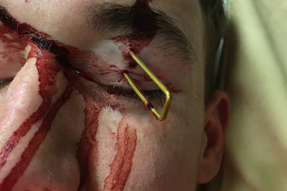
Penetrating ocular trauma, also known as an open globe injury, is a serious injury that involves an entry wound from the cornea or sclera. The injury is usually caused from a sharp and high velocity object (Figure 1A).

Figure 1 (A). Penetrating ocular trauma with nail foreign body present. (B). Ruptured globe injury.
Signs Of Penetrating Ocular Trauma
Specific signs will suggest a penetrating eye injury. A peaked or irregular pupil suggests a full thickness corneal laceration with loss of intraocular contents.
Examination may show a shallow anterior chamber in comparison to the other eye. Low intraocular pressure (IOP) should raise the suspicion of a ruptured globe (Figure 1B).
Haemorrhagic conjunctival chemosis (Figure 2) is highly suspicious of a globe rupture resulting from the escape of intraocular contents and blood. Urgent surgical exploration should be performed to exclude and repair the rupture.

Figure 2. Haemorrhagic conjunctival chemosis.
Case One
Hammering Metal
A 24-year-old construction worker presented with pain in the eye after hammering metal. He was not wearing eye protection. He also complained of severe redness and photophobia.
The visual acuity was hand movements and the IOP was not able to be assessed. External examination demonstrated a full thickness corneal laceration (Figure 3A) and a shallow anterior chamber with a developing lens opacity.
In this case it was important to image the posterior segment of the eye to exclude an IOFB. The CT scan demonstrated a metallic intraocular foreign body (Figure 3B).
Surgical repair of the anterior corneal wound, removal of the cataract, and a pars plana vitrectomy were performed to remove the metallic IOFB and repair the retinal damage (Figure 3C).

Figure 3. (A) Full thickness corneal laceration entry wound (arrow) in a hammering metal injury. (B) Computed tomography scan demonstrating a metallic foreign body in the posterior chamber. (C). Metallic foreign body in the posterior cavity during vitrectomy surgery. (D) IOFB measuring 0.5mm.
High Suspicion of IOFB
Proper history taking can provide clues of an IOFB. Metal hammering is an example of a high velocity injury that should provide a high index of suspicion for an IOFB, especially in cases where it may be difficult to identify an entry wound.
A foreign body can lodge into any ocular structure it encounters and travel into the globe. The consequences of missing the presence of an IOFB can be devastating. Stones and organic foreign bodies are associated with high infection rates. Therefore, close follow-up and thorough examination is often necessary.
Ocular imaging should always be considered in suspicious cases. An urgent CT scan of the orbits should be undertaken to rule out the presence of an IOFB. Magnetic resonance imaging (MRI) scans are contraindicated if there is suspicion of a metallic IOFB.
Case Two
Unusual Inflammation or Infection
A 40-year-old farmer presented with chronic intraocular inflammation. Two weeks previously he felt a ‘flick in the eye’ when cutting fencing wire. Shortly after, he was examined with no clear signs of ocular penetration.
Subsequently he developed persisting intraocular inflammation, which raised suspicion of an intraocular infection.
Visual acuity was 6/60 and IOP was 12mmHg. His conjunctiva was inflamed. Fine keratic precipitates with cells in the anterior chamber were noted. The vitreous was also inflamed.
A B-scan ultrasound and CT scan did not show an IOFB.
Vitrectomy surgery was performed (Figure 4A). An area of inflammatory abscess was noted in which eyelashes were embedded and removed. A sealed full thickness sclera laceration next to this was found repaired (Figure 4B). It was determined that the wire had flicked into the eye and pushed the eyelashes through a scleral wound and into the vitreous cavity (Figure 4C–D).
Figure 4. (A) Anterior segment photo during pars plana vitrectomy demonstrated severely injected conjunctiva. (B) Exploration demonstrating an old scleral wound. (C-D) Individual hairs identified in the posterior segment.
Missed IOFB can cause chronic endophthalmitis. Be suspicious of ocular trauma when eyes are inflamed or infected.
Metallic IOFB from high velocity injuries, such as hammering metal, are not usually associated with intraocular infection. However, the foreign body needs to be removed.
Trauma in an agricultural setting may be associated with a high risk of infection by virulent bacteria such as Bacillus cereus. Patients with this Bacillus endophthalmitis may be very unwell with high fever, proptosis, and significant visual loss.
Organic IOFB such as wood, vegetable matter, and in this case, eyelashes, are considered heavily contaminated and will cause severe and rapid onset of inflammation as well as high risk of infection. Severe inflammatory reactions can also be seen with foreign bodies composed of copper, vegetable matter, iron, steel, and tin. Less severe inflammatory reactions are seen with nickel, aluminium, mercury, and zinc.4
Endophthalmitis resulting from delays in treatment is often more severe than the initial injury. Early diagnosis and treatment of intraocular infection, removal of IOFB, and repair of the wound is essential.
Imaging Modalities to Identify IOFB
B-scan ultrasonography is a point-of-care test that can be helpful to identify an IOFB, particularly when there is a difficult view to the posterior segment. However, this is often not possible given the risk of causing further damage to the eye by causing extrusion of intraocular contents in severe penetrating eye injuries.
X-ray radiography can be helpful for localising the IOFB. It can identify radio-opaque foreign bodies, but it is not possible to detect radiolucent foreign bodies, such as wood or glass.

Figure 5. Plain film x-ray demonstrating a metallic screw within the orbit.
Computed tomography (CT scan) is the imaging modality of choice and considered the gold standard to identify the presence of an IOFB. A fine cut (1mm) CT scan should be performed to detect a small IOFB.

Figure 6. Computed tomography scan demonstrating a BB pellet in the posterior segment of the globe.
MRI is used in very selected situations due to its contraindication in the presence of injuries that involve metallic and magnetic materials. The risk of the magnetic field causing the IOFB to migrate, causing further damage to the intraocular tissue, could potentially be devastating. This imaging modality can be considered in cases where a CT scan or B-scan ultrasonography has not demonstrated an IOFB, but there is a high suspicion of a non-metallic IOFB.
First-Aid Management of Penetrating Eye Injuries
The eye should be protected with a rigid shield and not patched to avoid pressure that may expulse intraocular contents through the wound. Tetanus status must be checked and a booster given if not up to date. Broad spectrum antibiotics should be commenced, particularly in cases with suspected organic matter with high risk of endophthalmitis.
The patient should be fasted in the event of possible surgery and should present to the closest tertiary ophthalmic centre for specialist vitreoretinal surgical care as required.
Blunt Ocular Trauma
A blunt force injury may rupture the wall of the globe. The most common causes of blunt trauma are sporting injuries or assault.3 The injuries would include vitreous haemorrhage, retinal detachment, choroidal rupture, macular hole, optic nerve avulsion, and dislocated lens.
The identification of a choroidal rupture may be hidden at the onset of injury due to overlying blood
Case Three
Bruising of the Retina A 30-year-old male presented to the emergency department after a soccer ball kicked to the eye.
On examination his visual acuity was 6/7.5 and his IOP was 12mmHg. There was periorbital ecchymosis. His extra ocular movements were full. His anterior segment examination demonstrated a temporal subconjunctival haemorrhage. His posterior segment examination demonstrated a temporal area of retinal whitening, consistent with commotio retinae (Figure 7).

Figure 7. Commotio retinae. Acute retinal whitening in the temporal region.
Commotio Retinae
Commotio retinae is oedema and bruising of the neural retina due to blunt trauma to the globe causing a contrecoup injury. The shock wave from the force travels posterior and causes a concussive injury to the photoreceptor outer segments, resulting in a confluent area of retinal whitening. It most frequently affects the temporal fundus.
Patients can describe a scotoma, but more often are asymptomatic. In mild cases, no treatment is required, and the scotoma will resolve spontaneously over weeks. Close monitoring is necessary for the development of retinal detachments. In severe cases, progressive retinal pigmentary degeneration can occur from retinal pigment epithelium (RPE) cell hyperplasia and intraretinal migration.
There is usually no treatment required as it is a self-limiting condition unless there is significant foveal involvement. Patients should be educated on symptoms of retinal detachment and a repeat dilated fundus exam should be completed in a few weeks.
Case Four
Vitreous Haemorrhage and Retinal Detachment
A 40-year-old truck driver presented to the optometrist with worsening vision. He was seen one week prior, initially with a shadow in the nasal peripheral visual field. This was noted after he fell onto a door handle with direct impact to the globe. He was seen initially with a visual acuity of 6/12 and was diagnosed to have a vitreous haemorrhage secondary to a traumatic vitreous detachment.
The shadow enlarged and, over the course of the week, his vision deteriorated to hand motions in the left eye with an intraocular pressure of 14mmHg. His posterior segment examination demonstrated a giant retinal tear from the superotemporal quadrant (Figure 8).

Figure 8. Fundus photo of a delayed diagnosis of a traumatic giant retinal tear.
Traumatic Vitreous Detachment
A vitreous haemorrhage (VH) is most commonly seen due to a traumatic vitreous detachment.5 The presence of a VH raises concern of an underlying retinal tear, detachment, or other serious posterior segment injury. A careful dilated fundus examination or, in cases of difficult view, a B-scan ultrasound should be undertaken to rule this out. The patient should have close follow-up and should be educated on the symptoms of a retinal detachment.
Retinal Tears and Detachment
Trauma accounts for 10% of causes of all retinal detachments.3 This results from retinal dialysis, retina tears related to traumatic posterior vitreous detachments, and tears associated with commotio retinae.
Retinal dialysis (disinsertion of the retina at the ora serrata) may lead to traumatic retinal detachment. This is caused by a break at the ora serrata due to the traction from the inelastic vitreous gel along the posterior aspect of the vitreous base. It is most commonly seen in the superonasal and inferotemporal quadrants.
Retinal tears are another predisposing cause of a traumatic retinal detachment. The mechanism is due to abrupt deformation of the globe and equatorial expansion. The tears result from the strong vitreoretinal adhesions in the peripheral retina.
Trauma related to commotio retinae can also predispose to a retinal detachment within a few weeks of injury.
If retinal tears or detachments are identified, they should be urgently referred to a vitreoretinal service for repair to save sight and prevent long-term vision loss.
Case Five
Central Vision Loss
A 19-year-old student presented to the emergency department after sustaining an injury from a blackboard eraser hitting his eye. He complained of ocular pain and decreased vision.
On examination his visual acuity was 6/60 and his intraocular pressure was 12mmHg. His anterior segment examination was unremarkable. His posterior segment examination demonstrated a full thickness macular hole (Figure 9). An OCT was performed demonstrating a full thickness neuroretinal defect.

Figure 9. Fundus photo of a traumatic thickness macular hole.
Traumatic Macular Hole
A full thickness macular hole at or near the centre of the fovea can result from blunt trauma. The exact mechanism of traumatic macular holes is not well understood. Traumatic compression and expansion of the globe produces significant stress on the retina at the vitreoretinal interface causing separation. It is thought that it may develop in association with commotio retinae, or subretinal haemorrhage from a choroidal rupture.6
There is a higher likelihood of spontaneous closure of traumatic macular holes when compared to idiopathic macular holes.7 In cases where conservative management fails, pars plana vitrectomy with gas tamponade is an effective treatment, with satisfactory postoperative visual acuity.
Case Six
Sling Shot Injury
A 30-year-old presented to the emergency department after sustaining a sling-shot injury to the eye. He did not have any visual complaints other than minimal pain around the orbit.
On examination his visual acuity was 6/6 and his IOP was 14mmHg. His anterior segment examination was unremarkable. His posterior segment examination demonstrated a white crescent-shaped streak concentric to the optic nerve (Figure 10A).
An OCT scan demonstrated the loss of continuity of the RPE, with thinning of the underlying choroid (Figure 10C). A fluorescein angiogram was performed demonstrating expanding hyperfluorescence in the later phases of the study, which is consistent with choroidal neovascularization (Figure 10B).

Figure 10. (A) Fundus photo of the posterior segment showing a concentric crescent of a choroidal rupture. (B) Fluorescein angiography demonstrating the hypofluorescence in choroidal neovascularisation. (C) OCT scan demonstrating loss of RPE continuity and choroidal neovascularisation.
Choroidal Rupture and Choroidal Neovascularisation
A rupture of the choroid, Bruch’s membrane, and RPE is a serious complication of closed-globe trauma. It is seen in 5% of all closed-globe injuries.8 It is caused by the sudden change in the globe geometry from a compressive force against a relatively non-elastic Bruch’s membrane, choroid, and RPE. The identification of a choroidal rupture may be hidden at the onset of injury due to overlying blood. After the absorption of blood, a yellow or white streak of exposed underlying sclera, concentric to the optic disc, may be present. The sclera remains intact due to the high tensile strength.
Serial follow-up should be undertaken for these patients as this is often missed in the first instance due to the haemorrhage obscuring the underlying choroid. The rare consequence of choroidal neovascularization may develop in the region of the rupture and may require treatment with intravitreal anti-vascular endothelial growth factor.
After injury, patients require ongoing long-term follow-up for traumatic complications, which include cataract, glaucoma, corneal decompensation, and retinal scarring
Case Seven
Eye Gouging Rugby Injury
A 21-year-old college student presented to the emergency department with acute vision loss immediately following finger gouging of his left eye while playing rugby.
On examination his visual acuity was perception of light and normal IOP. He had a relative afferent pupillary defect. There was significant periorbital ecchymosis. His anterior segment examination demonstrated a hyphaema. A dilated fundus examination demonstrated a poorly visible optic disc with preretinal and subretinal haemorrhage surrounding the optic disc (Figure 11).

Figure 11. Optic nerve avulsion with severe optic nerve injury and vascular compromise.10
Optic Nerve Avulsion
An avulsion of the optic nerve is a rare occurrence but has significant visual consequences. The clinical presentation after trauma would be immediate profound visual loss at the time of injury. The examination of the posterior segment would demonstrate an absence of the optic nerve head and varying severities of haemorrhage.
It is thought that the avulsion occurs from extreme rotation of the globe relative to the nerve; an abrupt increase in IOP, which displaces the optic nerve out of the scleral canal, or an increase in intraorbital pressure, which displaces the optic nerve posteriorly and the globe anteriorly.9 Visual prognosis depends on the degree of the avulsion, and there is no available treatment for an optic nerve avulsion.
Long-Term Consequences Of Ocular Trauma
After injury, patients require ongoing long-term follow-up for traumatic complications, which include cataract, glaucoma, corneal decompensation, and retinal scarring. Management of these may require the care of multidisciplinary ophthalmic specialities and considerations for long-term rehabilitation.
Early diagnosis and referral for vitreoretinal care improves visual prognosis in these complex cases
Traumatic Glaucoma And Cataracts
Traumatic glaucoma can be caused by angle recession due to the rupture of the face of the ciliary body and the trabecular meshwork. Elevated IOP is likely to occur depending on the degree of recession. Other causes of glaucoma include chronic inflammation and lens damage, as well as complications of retinal detachment repair if silicone oil has been used as a tamponade agent. These patients will require lifelong follow-up and regular IOP checks.
The formation of a traumatic cataract depends on the mechanism of the injury. If the lens capsule is ruptured, the cataract can have a rapid onset as the lens fibres are being disrupted by the contact with the aqueous humour.
Trauma to the lens can cause lens particle glaucoma, where lens cortex particles circulate in the anterior chamber, causing a pro-inflammatory effect and raised IOP. Subsequent cataract surgery may be complicated by zonular weakness or capsular damage.
Loss Of The Globe
Unfortunately, in cases of severe injury, enucleation or evisceration may need to be performed. This is considered a last resort. Every attempt should be made to salvage the globe during the primary repair. Removal of the eye may be discussed after primary repair surgery if there is no prospect of salvageable vision, and the eye is a liability to the patient in the future. This requires counselling of the patient regarding ongoing care, risks of treatment, and sympathetic ophthalmia. The patient should also understand the long-term prospects of rehabilitation and ocular prothesis.
Conclusion
When a patient presents with posterior segment injury, there must always be a suspicion of a penetrating and blunt trauma. Accurate history taking and examination, with appropriate imaging of the posterior segment will provide clues to blinding pathology such as ruptured globe, penetrating wounds, intraocular foreign bodies, severe infection, and retinal detachment.
Early diagnosis and referral for vitreoretinal care improves visual prognosis in these complex cases. In the acute setting, modern vitreoretinal surgical techniques are effective in repairing injuries to the posterior segment. These patients require long-term follow-up by a multidisciplinary team for potential blinding complications due to injury such as cataract, glaucoma, and late retinal scarring.
 Dr Lawrence Kwok MD MMed (Ophth) is an Ophthalmology Registrar at Sydney, a Senior Resident Medical Officer at Sydney Eye Hospital, and an Honorary Researcher at the Royal Victorian Eye and Ear Hospital.
 Professor Andrew Chang AM MBBS (Hons), PhD, FRANZCO, FRACS
Andrew Chang is a vitreoretinal surgeon and ophthalmologist. He holds academic appointments of Conjoint Professor in the Department of Surgery at the University of NSW and Clinical Associate Professor at the University of Sydney. He is the Head of Ophthalmology at the Sydney Eye Hospital. He is the Medical Director of Sydney Retina Clinic.
References
1. Long, J., Mitchell, R. Hospitalised eye injuries in New South Wales, Australia. The Open Epidemiology Journal.2009;2:1–7.
2.May, D.R., Kuhn, F.P., Morris, R.E., et al. The epidemiology of serious eye injuries from the United States Eye Injury Registry. Graefes Arch Clin Exp Ophthalmol.2000;238(2):153–7.
3. Salmon, J.F., Kanski’s clinical ophthalmology: a systematic approach. Ninth edition. ed. Edinburgh:
Elsevier; 2020.
4. Bagheri, N., Wajda, B., Calvo, C., Durrani, A., The Wills Eye Manual: Office and Emergency Room Diagnosis and Treatment of Eye Disease. 7ed. Hagerstown: Wolters Kluwer; 2016.
5. Spraul, C.W., Grossniklaus, H.E., Vitreous hemorrhage. Surv Ophthalmol. 1997;42(1):3–39.
6.Williams, D.F., Mieler, W.F., Williams, G.A., Posterior segment manifestations of ocular trauma. Retina. 1990;10 Suppl 1:S35–44.
7. Tang, Y.F., Chang, A., Campbell, W.G., et al. Surgical management of traumatic macular hole: Optical coherence tomography features and outcomes. Retina.2020;40(2):290–8.
8. Bellows, J.G., Observations on 300 consecutive cases of ocular war injuries. Am J Ophthalmol. 1947;30(3):309–23.
9. Chong, C.C., Chang, A.A., Traumatic optic nerve avulsion and central retinal artery occlusion following rugby injury.Clin Exp Ophthalmol. 2006;34(1):88–9.
10. Image reproduced with permission from Chong, C.C.,Chang, A.A., Traumatic optic nerve avulsion and central retinal artery occlusion following rugby injury. Clin Exp Ophthalmol. 2006;34(1):88–9.


