

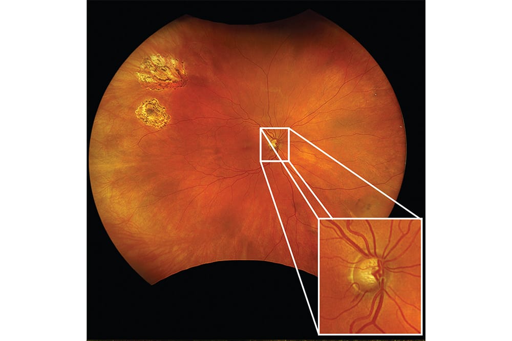
Optomap delivers detailed imaging from pole to periphery, allowing glaucoma specialists to effortlessly examine the entire posterior segment – even through a 2 mm pupil or a +3 dense cataract.
By incorporating optomap into examinations, specialists can confidently assess the entire retina, with studies showing a 30% increase in pathology detection compared to direct observation alone.1
With 97% of optomap users reporting the identification of unexpected pathologies in patients without visual complaints,2 this advanced technology empowers glaucoma specialists to provide superior glaucoma care and efficiently detect retinal diseases with precision.
Visit: optos.com
Reference
- Optos, Optomap increases percentage of pathology detected in conventional eye exam New England College of Optometry study published in Eye and Brain (media release, 15 April 2013) available at: optos.com/press-releases/optomap-increases-percentage-of-pathology-detected-in-conventional-eye-exam/ [accessed Feb 2025].


