Presbyopia can be regarded as the world’s largest refractive error, as almost every single human being on this planet will experience it from their 40s onwards.1 In 2019, as an ophthalmic surgeon establishing Perth’s first new refractive surgery practice in almost 20 years, Dr Lourens van Zyl quickly identified a significant market gap in the treatment of presbyopic patients; not just in Perth but across Australia.
When considering treatment options for presbyopia, several key criteria must be met for optimal outcomes. The ideal treatment should:
- Provide clear vision at near, intermediate, and distance ranges,
- Maintain excellent optical quality by preserving contrast sensitivity and nighttime vision,
- Preserve binocularity,
- Demonstrate high patient tolerance,
- Enable quick adaptation and return to normal activities,
- Simultaneously correct any refractive errors, and
- Offer reversibility if needed.
Currently, the most widely performed surgery for presbyopia worldwide is refractive lens exchange with the implantation of a diffractive multifocal intraocular lens (IOL).2-5 While lens extraction surgery is considered one of the safest operations in medicine,5,6 it remains an invasive procedure that carries risks of severe intraocular complications, including endophthalmitis, retinal detachments, and potential blindness.6,7 It’s worth noting that multifocal IOL implantation for presbyopia management is technically performed off-label, as these lenses are registered for cataract surgery rather than presbyopia treatment.2
Moreover, the implantation of a diffractive lens presents its own challenges. The human brain has not evolved to fully adapt to diffractive optics. Glare and haloes associated with these lenses never go away, despite patients being counselled that they will “get used to it”. This limitation often leads to lens exchange surgery when patients cannot adapt to the severe nighttime dysphotopsia of glare and haloes associated with diffractive IOLs.
Introducing Presbyond: A Sophisticated Solution
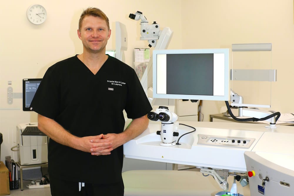
To differentiate my practice and address these challenges, I introduced Presbyond laser blended vision from ZEISS Meditec to Perth, after spending a significant amount of time training under its inventor, Professor Dan Reinstein. Presbyond represents a highly sophisticated treatment option for presbyopia that avoids the drawbacks of traditional surgical approaches.
Presbyond is a proprietary software developed by ZEISS Meditec for planning a customised binocular laser assisted in situ keratomileusis (LASIK) procedure that simultaneously manages presbyopia and corrects refractive error. The technology works by increasing the depth of field through controlled spherical aberration induction and creating a micro-anisometropia (≤1.5D). The procedure is performed using standard LASIK techniques.
Studies have shown that up to 97% of presbyopic patients can tolerate micro-anisometropia.6,8,9 What sets Presbyond apart is its ability to provide patients with a continuous range of quality vision from near to far, combining the accuracy of laser refractive procedures while avoiding common drawbacks like glare and haloes at nighttime. Importantly, patients treated with Presbyond maintain both stereoacuity and contrast sensitivity post-treatment.
Advanced Diagnostics
The sophistication of Presbyond treatment demands the most advanced diagnostic equipment for keratorefractive surgery. This led me to invest in two key pieces of technology: the CSO MS-39 anterior segment optical coherence tomography (OCT) with topographer, and the Osiris topographer and ocular aberrometer.
The MS-39: Comprehensive Corneal Analysis
The MS-39 combines anterior OCT with Placido disk corneal topography to generate high-resolution, detailed reports (Figures 1, 2).
Its capabilities include:
- Epithelial thickness and point spread function mapping,
- Early detection of corneal abnormalities through epithelial mapping and Gaussian reports,
- Built-in IOL calculations based on ray-tracing,
- LASIK flap thickness mapping,
- Detailed topography of both anterior and posterior cornea, and
- Keratoconus analysis function with Gaussian analysis.
The device’s ability to detect subclinical keratoconus through epithelial mapping is particularly valuable. Areas of epithelial thinning corresponding with increased elevation and tangential steepening are pathognomonic of early keratoconus.6-8 The MS-39 can also assess previous keratorefractive surgery cases where uncertainty exists.
The Osiris: Advanced Aberrometry
The Osiris complements the MS-39 by providing detailed corneal topography and higher-order aberration measurements across both the cornea and the whole eye (Figure 3). Its simulation maps facilitate patient education on complex visual problems. We combine the diagnostic output from both the MS-39 and Osiris T to not only assess the suitability of a patient to undergo Presbyond LASIK but also to give both a corneal and whole aberration profile that will be used by the Presbyond software to work out a bespoke plan for a patient.

Figure 1. Example of the MS-39 topography.

Figure 2. An example of an MS-39 corneal aberration profile.

Figure 3. Example of the Osiris T aberration profile printout with whole eye, corneal, and lenticular aberrations.
Understanding Spherical Aberrations in Presbyond
As Prebyond laser blended vision uses natural spherical aberrations, it is imperative to analyse and know what the corneal and whole eye aberration profile of the eye is. The Osiris T measures both whole eye and corneal Zernike higher order aberration profiles. The most important aberration is the Z(4,0) spherical aberration.
In most individuals, the cornea has positive spherical aberrations, which are counteracted by the negative spherical aberrations in the crystalline lens. To accurately perform Presbyond, the whole aberration profile is input in the software. This includes the aberration profile of the crystalline lens, which can counteract the induced spherical aberrations in myopic ablations or create toxic aberrations in hyperopic corrections. The human brain has evolved to filter spherical aberrations of up to either +0.6 µm or -0.6 µm, without affecting the quality of the image.8-10
The Presbyond Advantage: How It Works
Presbyond combines two key elements to achieve its results:
- Enhanced depth of field through controlled spherical aberrations, and
- Micro-anisometropia in the non-dominant eye.
The software induces spherical aberrations within tolerable ranges (less than ±0.6 µm) to enhance the depth of field by 1.5 diopters. As this alone isn’t sufficient for complete near to distance vision, a mild anisometropia is induced in the non-dominant eye, typically +0.75 to +1.5 diopters depending on the age of the patient and residual accommodative amplitude. In most cases the standard +1.5 diopters anisometropia in the non-dominant eye is sufficient,11-15 but as a rule we actively measure the residual accommodative amplitude of each patient to ensure a bespoke treatment that will be easy for a patient to adapt to.
Traditional monovision approaches using contact lenses or monofocal IOLs face several challenges including:
- Limited patient tolerance – not everyone can tolerate an anisometropia sufficient to offer reading and distance vision,
- Low patient satisfaction in many cases due to the compromises associated with monovision,
- Difficulty in accepting the near/distance vision discrepancy, summation, and loss of intermediate, especially where intermediate vision is essential for tablet and computer use, and
- Severely compromised depth perception in monovision.16-21
Presbyond overcomes these limitations by combining a micro-anisometropia with extended depth of focus induced by spherical aberrations to facilitate sharp, binocular vision from near over intermediate to far without compromising quality of vision. This solution achieves a 97% tolerance rate among presbyopic patients.6-8,19 The induced spherical aberrations remain at non-toxic levels, allowing the brain to filter them without compromising visual performance or contrast sensitivity.
The Blend Zone Advantage
Unlike standard monovision, which can create ‘blur zones’ requiring additional correction for intermediate tasks (Figure 4), Presbyond creates a ‘blend zone’ enabling crisp, continuous vision from near to far (Figure 5). The dominant eye maintains primary distance vision while gaining some near capability through enhanced spherical aberrations. Conversely, the non-dominant eye focusses primarily on near vision while retaining some distance capability.
This blend zone allows the brain to effectively fuse images from both eyes, maintaining binocularity. Non-dominant eyes targeted for -1.5D often achieve 6/7.5 or 6/9 unaided vision due to the enhanced depth of field, while dominant eyes can typically read N6 when aimed at emmetropia.6-8,19

Figure 4. In monovision there is a ‘blur-zone’ where a patient will still require glasses to see because the depth of field is not enhanced to cover that specific area of vision.

Figure 5. Anisometropia is induced in the non-dominant eye and there is an overlap of vision known as the ‘blend zone’.
|
||
|---|---|---|
| The MS-39 and Osiris aberrometer’s capabilities extend well beyond refractive surgery. For general ophthalmologists primarily performing cataract surgery, these devices offer exceptional diagnostic value that is often underutilised.
A neglected aspect of cataract surgery is the impact of corneal spherical aberrations on visual outcomes with specific lenses. I believe that corneal aberration profiles should be considered standard in cataract assessment, particularly in high-volume settings. High spherical aberrations can exacerbate nighttime glare and haloes with specific premium IOLs. Both negative and positive spherical aberrations above 0.6 µm can significantly impact contrast sensitivity. In cases where corneal aberrations make premium IOLs unsuitable, micro-monovision combined with existing spherical aberrations can offer an effective alternative for achieving spectacle independence. Understanding each IOL’s aberration profile allows surgeons to enhance visual outcomes by strategically working with patients’ existing corneal spherical aberrations. Managing KeratoconusThe MS-39 excels in monitoring progression following topographical trans-photorefractive keratectomy (PRK) treatment for irregular corneas, particularly in mild keratoconus cases treated with laser therapy. While conventional teaching advises against keratorefractive procedures on keratoconic corneas, in certain cases the Excimer laser is excellent in regularising a cornea. The MS-39 can be used and incorporated in the pre-op diagnostic workup of the Excimer laser to guide its surgery. In summary, the MS-39 is an invaluable tool when it comes to both corneal care as well as cataract surgeries in a general practice. |
References
- Karakus S. What is presbyopia, John Hopkins Medical Centre (webpage) avialable at: hopkinsmedicine.org/health/conditions-and-diseases/presbyopia#:~:text=Presbyopia%20eventually%20affects%20almost%20everyone,or%20nearsightedness%20along%20with%20presbyopia [accessed Feb 2025].
- Fricke TR, Tahhan N, Naidoo KS, et al. Global prevalence of presbyopia and vision impairment from uncorrected presbyopia: Systematic review, meta-analysis, and modelling. Ophthalmology. 2018;125(10):1492-9. doi: 10.1016/j.ophtha.2018.04.013.
- Wolffsohn JS, Davies LN. Presbyopia: Effectiveness of correction strategies. Prog Retin Eye Res. 2019;68:124-43. doi: 10.1016/j.preteyeres.2018.09.004.
- Glasser A, Campbell MC. Biometric, optical and physical changes in the isolated human crystalline lens with age in relation to presbyopia. Vision Res. 1999;39(11):1991-2015. doi: 10.1016/s0042-6989(98)00283-1.
- Patel I, West SK. Presbyopia: prevalence, impact, and interventions. Community Eye Health. 2007;20(63):40-1. PMID: 17971909.
- Reinstein DZ, Carp GI, Moore T, et al. LASIK for the correction of high hyperopic astigmatism with epithelial thickness monitoring. J Refract Surg. 2017;33(5):314-21. doi: 10.3928/1081597X-20170111-04.
- Vargas-Fragoso V, Alio JL. Corneal compensation of presbyopia: PresbyLASIK: an updated review. Eye Vis (Lond). 2017;4:11. doi: 10.1186/s40662-017-0075-9.
- Reinstein DZ, Archer TJ, Gobbe M. LASIK for myopic astigmatism and presbyopia using non-linear aspheric micro-monovision with the Carl Zeiss Meditec MEL 80 Platform. J Refract Surg. 2011;27(1):23-37. doi: 10.3928/1081597X-20100212-04.
- Reinstein DZ, Carp GI, Archer TJ, Gobbe M. LASIK for presbyopia correction in emmetropic patients using aspheric ablation profiles and a micro-monovision protocol with the Carl Zeiss Meditec MEL 80 and VisuMax. J Refract Surg. 2012;28(8):531-41. doi: 10.3928/1081597X-20120723-01.
- Reinstein DZ, Couch DG, Archer TJ. LASIK for hyperopic astigmatism and presbyopia using micro-monovision with the Carl Zeiss Meditec MEL80 platform. J Refract Surg. 2009;25(1):37-58. doi: 10.3928/1081597X-20090101-07.
- Schallhorn SC, Venter JA, Hannan SJ, Hettinger KA. Outcomes of wavefront-guided laser in situ keratomileusis using a new-generation Hartmann-Shack aberrometer in patients with high myopia. J Cataract Refract Surg. 2015;41(9):1810-9. doi: 10.1016/j.jcrs.2015.10.007.
- Alio JL, Grzybowski A, Romaniuk D. Refractive lens exchange in modern practice: when and when not to do it? Eye Vis (Lond). 2014;1:10. doi: 10.1186/s40662-014-0010-2.
- Lim DH, Chung ES, Kim MJ, Chung TY. Visual quality assessment after presbyopic laser in-situ keratomileusis. Int J Ophthalmol. 2018;11(3):462-9. doi: 10.18240/ijo.2018.03.17.
- Guzowski M, Wang JJ, Mitchell P, et al. Five-year refractive changes in an older population: the Blue Mountains Eye Study. Ophthalmology. 2003;110(7):1364-70. doi: 10.1016/S0161-6420(03)00465-2.
- Waring GO, 3rd. Standard graphs for reporting refractive surgery. J Refract Surg. 2000;16(4):459-66. PMID: 10939727.
- Paul C, Glaser S, Sekundo W, et al. Patient-reported quality of life and satisfaction after refractive lens extraction using a diffractive trifocal IOL: A multicenter retrospective cohort study. J Refract Surg. 2021;37(11):768-74. doi: 10.3928/1081597X-20210812-01.
- Artal P, Fernandez EJ, Manzanera S. Are optical aberrations during accommodation a significant problem for refractive surgery? J Refract Surg. 2002;18(5):S563-6. doi: 10.3928/1081-597X-20020901-13.
- Day AC, Donachie PH, Sparrow JM, Johnston RL, Royal College of Ophthalmologists’ National Ophthalmology D. The Royal College of Ophthalmologists’ National Ophthalmology Database study of cataract surgery: report 1, visual outcomes and complications. Eye (Lond). 2015;29(4):552-60. doi: 10.1038/eye.2015.3.
- Ganesh S, Brar S, Gautam M, Sriprakash K. Visual and refractive outcomes following laser blended vision using non-linear aspheric micro-monovision. J Refract Surg. 2020;36(5):300-7. doi: 10.3928/1081597X-20200407-02.
- Roberts C. The cornea is not a piece of plastic. J Refract Surg. 2000;16(4):407-13. doi: 10.3928/1081-597X-20000701-03.
- Zhang T, Sun Y, Liu Q, et al. Aspheric micro-monovision LASIK in correction of presbyopia and myopic astigmatism: Early clinical outcomes in a Chinese population. J Refract Surg. 2016;32(10):680-5. doi: 10.3928/1081597X-20160628-01.